Meiosis is a reduction division of a diploid nucleus to form haploid nuclei
Meiosis makes gametes (sex cells), which are produced in the reproductive organs (gonads).
4.2.2 Define homologous chromosomes
Homologous chromosomes resemble each other in structure. They occur in diploid cell, contain the same sequence of genes, but have come from different parents. They have the same genes at the same loci positions
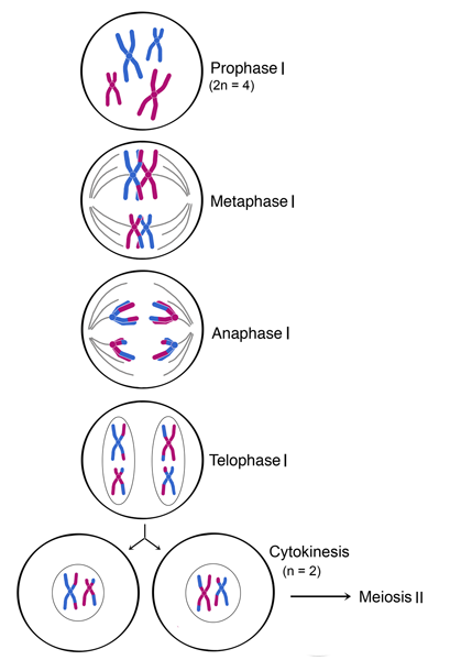
4.2.3 Outline the process of meiosis, including pairing of homologous chromosomes and crossing over, followed by two divisions, which results in four haploid cells
The process of meiosis involves two divisions, both of which follow the same basic stages as mitosis (prophase, metaphase, anaphase and telophase)
Meiosis is preceded by interphase, which includes the replication of DNA (S phase) to create chromosomes with genetically identical sister chromatids
Meiosis I
Homologous chromosomes must first pair up in order to be sorted into separate haploid daughter cells
In prophase I, homologous chromosomes undergo a process called synapsis, whereby homologous chromosomes pair up to forma bivalent (or tetrad)
- The homologous chromosomes are held together at points called chiasma (singular: chiasmata)
- Crossing over of genetic material between non-sister chromatids can occur at these points, resulting in new gene combination (recombination)
- Crossing over and independent assortment are the process which provides the genetic variation
The remainder of meiosis I involves separating the homologous chromosomes into separate daughter cells
- In meetaphase I, the homologous chromosomes pairs line up along the equator of the cell
- In anaphase I, the homologous chromosomes split apart and move to opposite poles
- In telophase I, the cell splits into two haploid daughter cells as cytokinesis happens concurrently
Meiosis II
In meiosis II, the sister chromatids are divided into separate cells
- In prophase II, spindle fibre reform and reconnect to the chromosomes
- In metaphase II, the chromosomes line up along the equator of the cell
- In anaphase II, the sister chromatids split apart and move to opposite poles
- In telophase II, the cell splits in two as cytokinesis happens concurrently.
Because sister chromatids may no longer be genetically identical as a result of potenial recombination, the process of meiosis results in the formation of four generally distinct haploid daughter cells
4.2.4 Explain that non-disjunction can lead to changes in chromosome number, illustrated by reference to Down syndrome (trisomy 21)
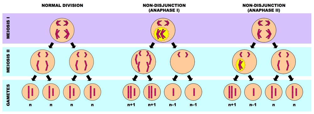
Non-disjunction refers to the chromosomes failing to separate correctly, resulting in gametes with one extra, or one missing chromosome (aneuploidy)
The failure of the chromosomes to separate may either occur via:
- Failure of homologous to separate during Anaphase I (resulting in four affected daughter cells)
- Failure of sister chromatids to separate during Anaphase II (resulting in two affected daughter cells)
Individuals with down syndrome has a trisomy 21 (three chromosomes 21)
4.2.5 State that, in karyotyping, chromosomes are arranged in pairs according to their size and structure
Karyotyping is a visual profile of all the chromosomes in a cell
The chromosomes are arranged into homologous pairs and displayed according to their structural characteristics
4.2.6 State that karyotyping is performed using cells collected by chorionic villus sampling or amniocentesis, for pre-natal diagnosis of chromosome abnormalities
Pre-natal karyotyping is often used to:
- Determine the gender of an unborn child (via identification of sex chromosomes)
- Test for chromosomal abnormalities (e.g. aneuploidies resulting from non-disjunction)
Amniocentesis
- A needle is inserted through the abdominal wall, into the amniotic cavity in the uterus, and a sample of amniotic fluid containing foetal cells is taken
- It can be done at ~ 16th week of pregnancy, with a slight chance of miscarriage (~0.5%)
Chorionic Villus Sampling
- A tube is inserted through the cervix and a tiny sample of the chronic villi (contains foetal cells) from the placenta is taken
- It can be done at ~ 11th week of pregnancy, with a slight risk of inducing miscarriage (~ 1%)
4.2.7 Analyse a human karyotype to determine gender and whether non-disjunction has occured.
Inside every cell in the human body, there are 46 chromosomes (except for red blood cells and haploid gametes)
Males have a X,Y chromosome while the female has X,X
Non-disjunction during gamete formation can lead to individuals with an abnormal number of chromosomes


沒有留言:
張貼留言