11.4.1 Annotate a light micrograph of testis tissue to show the location and function of interstitial cells (Leydig cells), germinal epithelium cells, developing spermatozoa and Sertoli cells
11.4.2 Outline the processes involved in spermatogenesis within the testis, including mitosis, cell growth, the two divisions of meiosis and cell differentiation
11.4.3 State the role of LH, testosterone and FSH in spermatogenesis
11.4.4 Annotate a diagram of the ovary to show the location and function of germinal epithelium, primary follicles, mature follicle and secondary oocyte
11.4.5 Outline the processes involved in oogenesis within the ovary, including mitosis, cell growth, the two divisions of meiosis, the unequal division of cytoplasm and the degeneration of polar body
11.4.6 Draw and label a diagram of a mature sperm and egg
11.4.7 Outline the role of the epididymis, seminal vesicle and prostate gland in the production of semen
11.4.8 Compare the processes of spermatogenesis and oogenesis, including the number of gametes and the timing of the formation and release of gametes
11.4.9 Describe the process of fertilization, including the acrosome reaction, penetration of the egg membrane by a sperm and the cortical reaction
11.4.10 Outline the role of HCG in early pregnancy
11.4.11 Outline the early embryo development up to the implantation of the blastocyst
11.4.12 Explain how the structure and functions of the placenta, including its hormonal role in the secretion of estrogen and progesterone, maintain pregnancy
11.4.13 State that the fetus is supported and protected by the amniotic sac and amniotic fluid
11.4.14 State that materials are exchanged between the maternal and fetal blood in the placenta
11.4.15 Outline the process of birth and its hormonal control, including the changes in progesterone and oxytocin levels and positive feedback
2014年6月9日 星期一
Topic 11.3: The kidney
11.3.1 Define excretion
11.3.2 Draw and label a diagram of the kidney
11.3.3 Annotate a diagram of a glomerulus and associated nephron to show the function of each part
11.3.4 Explain the process of ultrafiltration, including blood pressure, fenestrated blood capillaries and basement membrane
11.3.5 Define osmoregulation
11.3.6 Explain the reabsorption of glucose, water and salts in the proximal convulated tubule, including the roles of microvilli, osmosis and active transport
11.3.7 Explain the roles of the loop of Henle, medulla, collecting duct and ADH (vasopressin) in maintaining the water balance of the blood
11.3.8 Explain the differences in the concentration of proteins, glucose and urea between blood plasma, glomerular filtrate and urine
11.3.9 Explain the presence of glucose in the urine of untreated diabetic patients
Topic 11.2: Muscles and movement
11.2.1 State the roles of bones, ligaments, muscles, tendons and nerves in human movement
11.2.2 Label a diagram of the human elbow join, including cartilages, synovial fluid, joint capsule, named bones and antagonistic muscles (biceps and triceps)
11.2.3 Outline the functions of the structures in the human elbow joint named in 11.2.2
11.2.4 Compare the movements in the hip joint and the knee joint
11.2.5 Describe the structure of striated muscle fibres, including the myofibrils with light and dark bands, mitochondria, the sarcoplasmic reticulum, nuclei and the sacrolemma
11.2.6 Draw and label a diagram to show the structure of a sacromere, including Z lines, actin filaments, myosin filaments with heads, and the resultant light and dark bands
11.2.7 Explain how skeletal muscles contracts, including the release of calcium ions from the sacroplasmic reticulum, the formation of cross-bridges, the sliding of actin and myosin filaments, and the use of ATP to break cross-bridges and re-set myosin heads
11.2.8 Analyse electron micrographs to find the state of contraction of muscles fibres
Topic 11.1: Defense against infectious disease
11.1.1 Describe the process of blood clotting
11.1.2 Outline the principle of challenge and response, clonal selection and memory cells as the basis of immunity
11.1.3 Define active and passive immunity
11.1.4 Explain antibody production
11.1.5 Describe the production of monoclonal antibodies and their use in diagnosis and in treatment
11.1.6 Explain the principle of vaccination
11.1.7 Discuss the benefits and dangers of vaccination
Topic 11: Human health and physiology
Topic 11 of the IB HL Biology syllabus is the Genetics. IBO recommends to spend 17 hours on this topic.
This topic has 4 sub-chapters: "Defense against infectious disease", "Muscles and movement", "The kidney" and "Reproduction". Each are separated with numerical values in order of mentioned.
These are all HL syllabus statements, it is recommended to bring a Casio Graphical Calculator instead of Texas.
Topic 6.6: Reproduction
6.6.1 Draw and label diagrams of the adult male and female reproductive systems
6.6.2 Outline the role of hormones in the menstrual cycle, including FSH (follicle stimulating hormones), LH (luteinizing hormone), estrogen and progesterone
6.6.3 Annotate a graph showing hormone levels in the menstrual cycle, illustrating the relationship between changes in hormone level and ovulation, menstruation and thickening of the endometrium
6.6.4 List three roles of testosterone in males
6.6.5 Outline the process of in vitro fertilization (IVF)
6.6.6 Discuss the ethical issues associated with IVF
Topic 6.5: Nerves, hormones and homeostasis
6.5.1 State that the nervous system consists of the central nervous system (CNS) and peripheral nerves, and is composed of cells called neurons that can carry rapid electrical impulses
6.5.2 Draw and label a diagram of the structure of a motor neuron
6.5.3 State that nerve impulses are conducted from recpetors to the CNS by sensory neurons, within the CNS by relay neurons, and from the CNS to effectors by motor neurons
6.5.4 Define resting potential and action potential (depolarization and repolarization)
6.5.5 Explain how a nerve impulse passes along a non-myelinated neuron
6.5.6 Explain the principles of synaptic transmission
6.5.7 State that the endocrine system consists of glands that release hormones that are transported in the blood
6.5.8 State that homeostasis involves maintaining the internal environment between limits, including blood pH, carbon dioxide concentration, blood glucose concentration, body temperature and water balance
6.5.9 Explain that homeostasis involves monitoring levels of variables and correcting changes in levels by negative feedback mechanism
6.5.10 Explain the control of body temperature, including the transfer of heat in blood, and the roles of the hypothalamus, sweat glands, skin arterioles and shivering
6.5.11 Explain the control of blood glucose concentration, including the roles of glucagon, insulin and a and b cells in the pancreatic islets
6.5.12 Distinguish between type I and type II diabetes
Topic 6.4: Gas exchange
6.4.1 Distinguish between ventilation, gas exchange and cell respiration
6.4.2 Explain the need for a ventilation system
6.4.3 Describe the features of alveoli that adapt them to gas exchange
6.4.4 Draw and label a diagram of the ventilation system, including trachea, lungs, bronchi, bronchioles and alveoli
6.4.5 Explain the mechanism of ventilation of the lungs in terms of volume and pressure changes caused by the internal and external intercostal muscles, the diaphragm and abdominal muscles
Topic 6.3: Defense against infectious disease
6.3.1 Define pathogen
A pathogen is an organism or virus that causes a disease. Most, but not all, are microorganisms.

6.3.2 Explain why antibiotics are effective against bacteria but not against viruses
Antibiotics are naturally occuring substances that slow down or kill microorganisms. They are obtained mainly from fungi or bacteria, and are substances which such organisms manufacture in their natural habitats.
Most antibiotics are so extremely effective at disrupting bacterial metabolism that whole populations are quickly suppressed. A key process here is the formation and laying down of the new dividing wall. Bacterial cells have a rigid wall containing giant, complex, molecules of amino-sugars and peptide units (polymers) rather than the polysaccharides of plant cell walls.

Virus on the other hand, are not living cells and have no metabolism of their own to be interfered with. Viruses reproduce using metabolic pathways in their host cell that are not affected by antibiotics. Antibiotics cannot be used to prevent viral diseases.

6.3.3 Outline the role of skin and mucous membranes in defense against pathogens
6.3.4 Outline how phagocytes leucocytes ingest pathogens in the blood and in blood tissues
6.3.5 Distinguish between antigens and antibodies
6.3.6 Explain antibody production
6.3.7 Outline the effects of HIV on the immune system
6.3.8 Discuss the cause, transmission and social implications of AIDs
A pathogen is an organism or virus that causes a disease. Most, but not all, are microorganisms.
6.3.2 Explain why antibiotics are effective against bacteria but not against viruses
Antibiotics are naturally occuring substances that slow down or kill microorganisms. They are obtained mainly from fungi or bacteria, and are substances which such organisms manufacture in their natural habitats.
Most antibiotics are so extremely effective at disrupting bacterial metabolism that whole populations are quickly suppressed. A key process here is the formation and laying down of the new dividing wall. Bacterial cells have a rigid wall containing giant, complex, molecules of amino-sugars and peptide units (polymers) rather than the polysaccharides of plant cell walls.
Virus on the other hand, are not living cells and have no metabolism of their own to be interfered with. Viruses reproduce using metabolic pathways in their host cell that are not affected by antibiotics. Antibiotics cannot be used to prevent viral diseases.
6.3.3 Outline the role of skin and mucous membranes in defense against pathogens
6.3.4 Outline how phagocytes leucocytes ingest pathogens in the blood and in blood tissues
6.3.5 Distinguish between antigens and antibodies
6.3.6 Explain antibody production
6.3.7 Outline the effects of HIV on the immune system
6.3.8 Discuss the cause, transmission and social implications of AIDs
Topic 6.2: The transport system
6.2.1 Draw and label a diagram of the heart showing the four chambers, associated blood vessels, valves and the route of blood through the heart

6.2.2 State that the coronary arteries supply heart muscle with oxygen and nutrients
The heart is a muscle itself, so note that the walls of the heart are supplied with oxygenated blood via coronary arteries. These arteries and the capillaries they serve, deliver to the muscle fibres of the heart the oxygen and the nutrients essential for the pumping action.

6.2.3 Explain the action of the heart in terms of collecting blood, pumping blood, and opening and closing of valves
The cavity of the heart is divided into four chambers, with those on the right side of the heart completely separate from those on the left. The two upper chambers are called the artia and these receive blood into the heart.
The lower chambers are thick-walled ventricles, with the muscular wall of the left ventricle much thicker than that of the right ventricle. However, the volumes of the right and left sides (the quantities of blood they contain) are identical. The ventricles pump blood out of the heart.
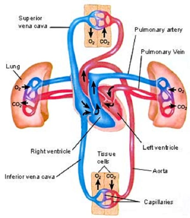
The valves of the heart prevent backflow of the blood, thereby maintaining the direction of flow through the heart. The artio-ventricular valves are large valves, positioned to prevent backflow from ventricles to artia. The edges of these valves are supported by tendons anchored to the muscle walls of the ventricles below. A different type of valve separates the ventricles from the pulmonary artery and the aorta. These are called the semi-lunar valves.

6.2.4 Outline the control of the heartbeat in terms of myogenic muscle contraction, the role of the pacemaker, nerves, the medulla of the brain and epinephrine (adrenaline)
The heart normally beats about 75 times per minute - approximately 0.8 seconds per beat. In each beat, the heart muscle contracts strongly, followed by a period of relaxation.
The atrium contacts pushing blood past the bicuspid valve into the ventricle, Atrium muscle relax. Ventricle muscles contract, causing blood pressure to close the bicuspid valve and open the semilunar valves, forcing blood into the aorta. Ventricle and atrium muscle relax, while the pressure of blood in the aorta causes the semilunar valves to shut. Blood flows into the atrium and opens the bicuspid valve as it starts to flow into the ventricle.
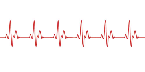 The heart beats rhythmically throughout life, without rest, apart from the momentary relaxation between beats. Even more remarkably, the origin of each beat is within the heart itself - we say the heart beat is myogenic in origin.
Beats originate in a strucuture in the muscle of the wall of the right atrium, called the pacemaker. Special muscle fibre radiate out from the pacemaker, conducting the impulse to the muscles of both artia, triggering contraction. Then a second excitation is passed onto the ventricles causing a ventricular contraction.

The heart receives impulses from a control centre in the hindbrain (medulla), via two nerves. One nerve, when stimulated, triggers speeding up of the heart rate, and the other nerve triggers a slowing down of the heart. Since these nerves have opposite effect, they are antagonistic.

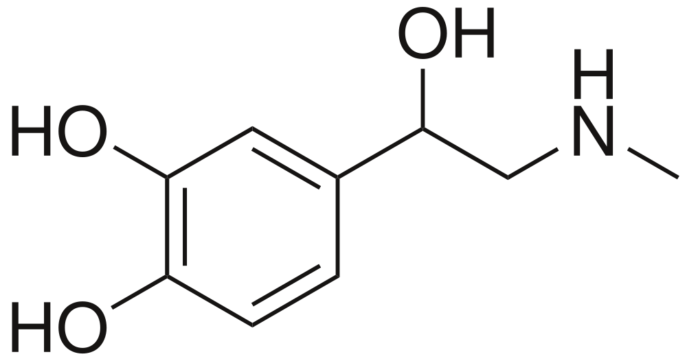
The hormone adrenaline is secreted by the adrenal glands and carried in the blood, causes the pacemaker to increase the heart rate.
6.2.5 Explain the relationship between the structure and function of arteries, capillaries and veins
There are three types of vessels in the circulation system.
Arteries
- Carry blood away from the heart
- Carry blood at high pressures
- Have a narrow lumen
- Have a thick middle layer to help the pulse flow
- Have a outer layer to prevent rupture
Capillaries
- Are a fine networks of tiny tubes linking arteries and veins.
- Involved with material and gas exchange with the surrounding body tissues
- Blood pressure is relatively low
- Small diametre
- Single cell to allow easier diffusion
- Contains pores to aid the transport of materials
Veins
- Carry blood to the heart
- Carry blood at low pressure
- Very wide lumen
- Have valves to prevent blooding pooling
- Thin surrounding tissue as blood do not go in rhythmic pulse.

6.2.6 State that blood is composed of plasma, erythrocytes, leucocytes (phagocytes and lymphocytes) and platelets
Blood is a special tissue consisting of a liquid medium called plasma in which are suspended red cells or erythrocytes, white cells or luecocytes, and platelets. The plasma is the medium for exchange of substances between cells and tissues, the erythrocytes are involved in transport of respiratory gases.

6.2.7 State that the following are transported by the blood: nutrients, oxygen, carbon dioxide, hormones, antibodies, urea and heat
The blood circulation has roles in the body's defense against diseases as well as being the all-important transport system of the body. Nutrients from digestion, oxygen and carbon dioxide, urea, hormones and antibodies are all transported. In the tissues of the body, exchange between the blood and cells of the tissues occurs from the capillaries, the walls of which are permeable and highly "leaky".
Tissue respiration - gas exchange
Hydration - water to all the tissues
Nutrition - Nutrients (sugars, amino acids, lipids, vitamins) and inorganic ions to all cells.
Excretion - Waste product urea to kidneys
Development and co-ordination - Hormones from endocrine glands to target organs
Temperature regulation - distribution of heat
Defense against disease - Antibodies are circulated in the blood system

6.2.2 State that the coronary arteries supply heart muscle with oxygen and nutrients
The heart is a muscle itself, so note that the walls of the heart are supplied with oxygenated blood via coronary arteries. These arteries and the capillaries they serve, deliver to the muscle fibres of the heart the oxygen and the nutrients essential for the pumping action.
6.2.3 Explain the action of the heart in terms of collecting blood, pumping blood, and opening and closing of valves
The cavity of the heart is divided into four chambers, with those on the right side of the heart completely separate from those on the left. The two upper chambers are called the artia and these receive blood into the heart.
The lower chambers are thick-walled ventricles, with the muscular wall of the left ventricle much thicker than that of the right ventricle. However, the volumes of the right and left sides (the quantities of blood they contain) are identical. The ventricles pump blood out of the heart.
The valves of the heart prevent backflow of the blood, thereby maintaining the direction of flow through the heart. The artio-ventricular valves are large valves, positioned to prevent backflow from ventricles to artia. The edges of these valves are supported by tendons anchored to the muscle walls of the ventricles below. A different type of valve separates the ventricles from the pulmonary artery and the aorta. These are called the semi-lunar valves.
6.2.4 Outline the control of the heartbeat in terms of myogenic muscle contraction, the role of the pacemaker, nerves, the medulla of the brain and epinephrine (adrenaline)
The heart normally beats about 75 times per minute - approximately 0.8 seconds per beat. In each beat, the heart muscle contracts strongly, followed by a period of relaxation.
The atrium contacts pushing blood past the bicuspid valve into the ventricle, Atrium muscle relax. Ventricle muscles contract, causing blood pressure to close the bicuspid valve and open the semilunar valves, forcing blood into the aorta. Ventricle and atrium muscle relax, while the pressure of blood in the aorta causes the semilunar valves to shut. Blood flows into the atrium and opens the bicuspid valve as it starts to flow into the ventricle.
Beats originate in a strucuture in the muscle of the wall of the right atrium, called the pacemaker. Special muscle fibre radiate out from the pacemaker, conducting the impulse to the muscles of both artia, triggering contraction. Then a second excitation is passed onto the ventricles causing a ventricular contraction.
The heart receives impulses from a control centre in the hindbrain (medulla), via two nerves. One nerve, when stimulated, triggers speeding up of the heart rate, and the other nerve triggers a slowing down of the heart. Since these nerves have opposite effect, they are antagonistic.
The hormone adrenaline is secreted by the adrenal glands and carried in the blood, causes the pacemaker to increase the heart rate.
6.2.5 Explain the relationship between the structure and function of arteries, capillaries and veins
There are three types of vessels in the circulation system.
Arteries
- Carry blood away from the heart
- Carry blood at high pressures
- Have a narrow lumen
- Have a thick middle layer to help the pulse flow
- Have a outer layer to prevent rupture
Capillaries
- Are a fine networks of tiny tubes linking arteries and veins.
- Involved with material and gas exchange with the surrounding body tissues
- Blood pressure is relatively low
- Small diametre
- Single cell to allow easier diffusion
- Contains pores to aid the transport of materials
Veins
- Carry blood to the heart
- Carry blood at low pressure
- Very wide lumen
- Have valves to prevent blooding pooling
- Thin surrounding tissue as blood do not go in rhythmic pulse.
6.2.6 State that blood is composed of plasma, erythrocytes, leucocytes (phagocytes and lymphocytes) and platelets
Blood is a special tissue consisting of a liquid medium called plasma in which are suspended red cells or erythrocytes, white cells or luecocytes, and platelets. The plasma is the medium for exchange of substances between cells and tissues, the erythrocytes are involved in transport of respiratory gases.
6.2.7 State that the following are transported by the blood: nutrients, oxygen, carbon dioxide, hormones, antibodies, urea and heat
The blood circulation has roles in the body's defense against diseases as well as being the all-important transport system of the body. Nutrients from digestion, oxygen and carbon dioxide, urea, hormones and antibodies are all transported. In the tissues of the body, exchange between the blood and cells of the tissues occurs from the capillaries, the walls of which are permeable and highly "leaky".
Tissue respiration - gas exchange
Hydration - water to all the tissues
Nutrition - Nutrients (sugars, amino acids, lipids, vitamins) and inorganic ions to all cells.
Excretion - Waste product urea to kidneys
Development and co-ordination - Hormones from endocrine glands to target organs
Temperature regulation - distribution of heat
Defense against disease - Antibodies are circulated in the blood system
Topic 6.1: Digestion
6.1.1 Explain why digestion of large food molecules is essential.
Most food is solid and in the form of large complex molecules which are insoluble and chemically inert (not readily stable)
Large molecules need to be broken down into smaller molecules that can be readily absorbed across membranes and into cells. Small molecules can be reassembled into new products (e.g. amino acids can be reassembled to form new proteins).

6.1.2 Explain the need for enzymes in digestion
Enzymes are biological catalysts that speeds up the rate of reaction. It allows reactions to take place at body temperatures. They are specific for a certain type of reaction though based on the lock and key model.

6.1.3 State the source, substrate, products and optimum pH conditions for one amylase, one protease and one lipase
Amylase
Example - Salivary amylase
Source - Salivary glands
Substrate - Starch
Product - Maltose
Optimum pH - 7
Protease
Example - Pepsin
Source - Gastric juice
Substrate - Protein
Product - Short polypeptides
Optimum pH - 2
Lipase
Example - Pancreatic juice
Source - Pancreas
Substrate - Lipids (triglyceride)
Product - Glycerol and fatty acids
Optimum pH - 8
6.1.4 Draw and label a diagram of the digestive system

6.1.5 Outline the function of the stomach, small intestine and large intestine
Stomach
Present in the wall of the stomach are millions of tiny pits called gastric glands which secrete the components of gastric juice. This juice includes hydrochloric acid - sufficiently acidic to create an environment of pH 1.5-2.0, which is the optimum pH for protein digestion by the protease enzymes of the gastric juice. These proteases, of which pepsi is one, are formed in cells of gastric glands and secreted in an inactive state. The hydrochloric acid then activates them, and kills most of the in coming bacteria in the food.
The whole stomach lining is supplied with goblet cells that secrete mucus. Mucus bathes the interior lining of the stomach, forming an effective barrier to both the hydrochloric acid and the protease of the gastric juices, preventing autolysis (self-digestion) of the stomach wall.
As the food is mixed with gastric juice and churned by muscle action it becomes a semi-liquid called chyme. The churning action of the stomach is an important part of the mechanical digestion process. A typical meal may spend up to four hours in the stomach.

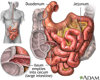
Small intestines
Food enters the first part of the small intestine (known as the duodenum) a little at a time. Here the chyme meets bile from the bile duct, and the pancreatic juice from the pancreas. Bile is strongly alkaline and neutralises the acidity of the chyme. It also lowers the surface tension of large fat globules, causing them to break into tiny droplets, a process called emulsification. This speeds digestion by the enzyme lipase later on. Bile itself contains no enzyme.
All these enzymes act as the chyme, bile and pancreatic juice are mixed together by a churning action (a form of peristalsis) called segmentation.
6.1.6 Distinguish between absorption and assimilation
Absorption can be defined as the movement of particles or dissolved substances across a membrane
Assimilation can be defined as the conversion of nutrients into fluids or solid parts of an organism.

6.1.7 Explain how the structure of the villus is related to its role in absorption and transport of the products of digestion
There are different structure of the villus which aid its role in absorption.
Micro-villi is present to provide a huge surface area for absorption.
Epithelium cells has a single layer of small cells, packed with mitochondria - the source of ATP for active uptake across the plasma membrane.
Protein pumps in the plasma membrane of the epithelial cells can actively transport nutrients across the plasma membrane into the villi
There is also a big network of capillaries which proved a large surface area for uptake of amino acids, monosaccharides, and fatty acids and glycerol into blood circulation.
Lacteal is a branch of the lymphatic system into which triglycerides (combined with protein) pass for transport to body cells
The mucus from goblet cells in the epithelium can lubricate movement of digested food among the villi and protects plasma membrane of epithelial cells.

Most food is solid and in the form of large complex molecules which are insoluble and chemically inert (not readily stable)
Large molecules need to be broken down into smaller molecules that can be readily absorbed across membranes and into cells. Small molecules can be reassembled into new products (e.g. amino acids can be reassembled to form new proteins).
6.1.2 Explain the need for enzymes in digestion
Enzymes are biological catalysts that speeds up the rate of reaction. It allows reactions to take place at body temperatures. They are specific for a certain type of reaction though based on the lock and key model.
6.1.3 State the source, substrate, products and optimum pH conditions for one amylase, one protease and one lipase
Amylase
Example - Salivary amylase
Source - Salivary glands
Substrate - Starch
Product - Maltose
Optimum pH - 7
Protease
Example - Pepsin
Source - Gastric juice
Substrate - Protein
Product - Short polypeptides
Optimum pH - 2
Lipase
Example - Pancreatic juice
Source - Pancreas
Substrate - Lipids (triglyceride)
Product - Glycerol and fatty acids
Optimum pH - 8
6.1.4 Draw and label a diagram of the digestive system
6.1.5 Outline the function of the stomach, small intestine and large intestine
Stomach
Present in the wall of the stomach are millions of tiny pits called gastric glands which secrete the components of gastric juice. This juice includes hydrochloric acid - sufficiently acidic to create an environment of pH 1.5-2.0, which is the optimum pH for protein digestion by the protease enzymes of the gastric juice. These proteases, of which pepsi is one, are formed in cells of gastric glands and secreted in an inactive state. The hydrochloric acid then activates them, and kills most of the in coming bacteria in the food.
The whole stomach lining is supplied with goblet cells that secrete mucus. Mucus bathes the interior lining of the stomach, forming an effective barrier to both the hydrochloric acid and the protease of the gastric juices, preventing autolysis (self-digestion) of the stomach wall.
As the food is mixed with gastric juice and churned by muscle action it becomes a semi-liquid called chyme. The churning action of the stomach is an important part of the mechanical digestion process. A typical meal may spend up to four hours in the stomach.
Small intestines
Food enters the first part of the small intestine (known as the duodenum) a little at a time. Here the chyme meets bile from the bile duct, and the pancreatic juice from the pancreas. Bile is strongly alkaline and neutralises the acidity of the chyme. It also lowers the surface tension of large fat globules, causing them to break into tiny droplets, a process called emulsification. This speeds digestion by the enzyme lipase later on. Bile itself contains no enzyme.
All these enzymes act as the chyme, bile and pancreatic juice are mixed together by a churning action (a form of peristalsis) called segmentation.
6.1.6 Distinguish between absorption and assimilation
Absorption can be defined as the movement of particles or dissolved substances across a membrane
Assimilation can be defined as the conversion of nutrients into fluids or solid parts of an organism.
6.1.7 Explain how the structure of the villus is related to its role in absorption and transport of the products of digestion
There are different structure of the villus which aid its role in absorption.
Micro-villi is present to provide a huge surface area for absorption.
Epithelium cells has a single layer of small cells, packed with mitochondria - the source of ATP for active uptake across the plasma membrane.
Protein pumps in the plasma membrane of the epithelial cells can actively transport nutrients across the plasma membrane into the villi
There is also a big network of capillaries which proved a large surface area for uptake of amino acids, monosaccharides, and fatty acids and glycerol into blood circulation.
Lacteal is a branch of the lymphatic system into which triglycerides (combined with protein) pass for transport to body cells
The mucus from goblet cells in the epithelium can lubricate movement of digested food among the villi and protects plasma membrane of epithelial cells.
Topic 6: Human health and physiology
Topic 6 of the IB SL Biology syllabus is the Human health and physiology. IBO recommends to spend 20 hours on this topic.
This topic has 6 sub-chapters: "Digestion", "The transport system", "Defense against infectious defense", "Gas exchange", "Nerves, hormones and homeostasis" and "Reproduction". Each are separated with numerical values in order of mentioned.
These are all SL syllabus statements, it is recommended to bring a Casio Graphical Calculator instead of Texas.
訂閱:
意見 (Atom)