2013年9月15日 星期日

Topic 2.5: Cell Division

2.5.1 Outline the stages in the cell cycle including interphases (G1, S, G2)


Interphase consists of G1, S and G2.

G1 is the first growth phase. Cytoplasm are active and new organelles are formed. It is also a phase of intense biochemical activity of growing cells

S is the synthesis of DNA, this is the phase here chromosomes are copied or replicated. The process in which chromatids

G2 is the second growth phase. It prepares the cell to undergo mitosis by increasing the growth

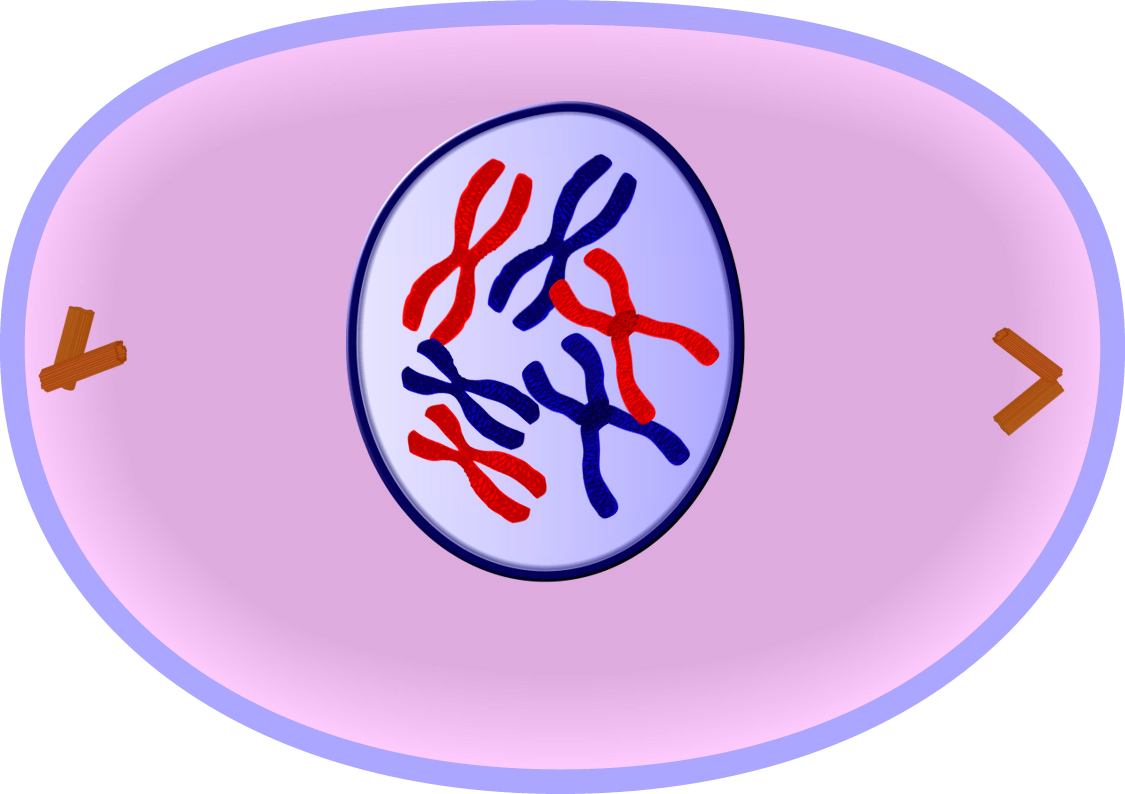
In prophase, chromosomes become visible as long thin threads. At the end of prophase, it is possible to see two chromatids held together at the centromere. The nuclear membrane starts the break down.

In metaphase, the centrioles move to opposite ends of the cell. Microtubules in the cytoplasm starts to form into a spindle attached to the centromeres of each pair of chromatids at the equator.


In anaphase, the centromeres divide, the spindle fibre shorten, and the chromatids are pulled by their centromeres to opposite poles.

In telophase, the nuclear membrane reforms and the chromosomes decondense by uncoiling. They become chromatin again.

Cytokinesis follows telophase where it splits the cell into two daughter cells.


2.5.2 State that tumours (cancers) are the result of uncontrolled cell division and that these can occur in any organ or tissue.

Uncontrolled cell division are various forms of different cancers affecting several tissues of the body. In cancer, cells divide by mitosis repeatedly, without control or regulation, forming an irregular mass of cells, called a tumour. Cancer is caused by damage to DNA of chromosomes. Mistakes of different types build up in the DNA of the body cells.


2.5.3 State that interphase is an active period in the life of a cell when many metabolic reactions occur, including protein synthesis, DNA replication and an increase in the number of mitochondria and/or chloroplast.

Interphase is an active period in the life of a cell during which many metabolic reactions occur such as protein synthesis, DNA replication and an increase in the number of mitochondria and/or chloroplast.


2.5.4 Describe the events that occur in the four phases of mitosis (prophase, metaphase, anaphase and telophase).


Prophase


Metaphase

Anaphase


Telophase


2.5.5 Explain how mitosis produces two genetically identical nuclei.

Daughter cells produced by mitosis have a set of chromosomes identical to each other and to the parent cell from which they were formed. This occurs because:

  • An exact copy of each chromosomes is made by accurate replication during interphase, when two chromatids are formed
  • Chromatids remain attached by their centromeres during metaphase of mitosis, when each becomes attached to a spindle fibre at the equator of the spindle
  • Centromeres then divide during anaphase and the chromatids of each pairs are pulled apart to opposite poles of the spindle.
  • The chromosomes at the poles form the new nuclei.
  • Two cells are then formed by division of the cytoplasm at the midpoint.


2.5.6 State that growth, embryonic development, tissue repair, and asexual reproduction involve mitosis.

In growth and development of an embryo, it is important that all cells carry the same genetic information as the existing cells from which they are formed, and which they share with surrounding cells or tissues. Similarly, when repair of damaged or worn out cells occurs, they are exact copies what they replace. In fact, this essential for growth, development and repair, because otherwise different parts of our body might start working to conflicting blueprints. The results will be chaos.

Mitosis cell division is also the basis of all forms of asexual reproduction, in which the offspring produced are identical to the parent.


Topic 2.4: Membranes

2.4.1 Draw and label a diagram to show the structure of membranes. (The diagram should show the phospholipid bilayer, cholesterol, glycoproteins, and integral and peripheral proteins.)

The plasma membrane is made almost entirely of protein and lipid,together with a small and variable amount of carbohydrate. This is called the fluid mosaic model, it is described as fluid because the components (lipids and proteins) are on the move and mosaic because the proteins are scattered about in this pattern.


All cells have a plasma/cell membrane

Functions
  • Controls what enters and exits the cell to maintain and internal balance called homeostasis
  • Provides protection and support for the cell
Structure (Phospholipid layer)
  • Phosphate head is polar (water loving)
  • Fatty acid tails non-polar (water fearing)
  • Protein embedded in membrane
They are also selectively permeable: allows some molecules in and keeps other molecules out


2.4.2 Explain how the hydrophobic and hydrophilic properties of phospholipids help to maintain the structure of cell membranes.

Phospholipid has a head composed of a glycerol group to which is attached to one ionized phosphate group. This latter part of the molecule has hydrophilic properties (water-loving). Hydrogen bonds form between the phosphate head and the water molecules.

The remainder of the phospholipid consists of two long, fatty acid residues consisting of hydrocarbon chains. These 'tails' have hydrophobic properties (water-hating).

Therefore phospholipid is unusual in being partly hydrophilic and partly hydrophobic.

When more phospholipid is available, the molecules arrange themselves as a bi-layer, with the hydrocarbon tails facing together. In the lipid bi-layer, attractions between the hydrophobic hydrocarbon tails are on the inside which the hydrophilic heads are on the outside. The surrounding water on the outside make a stable and strong barrier.


2.4.3 List the functions of membrane proteins.

Movement of molecules across the plasma membrane of living cells is continuous and heavy. Cells requires the plasma membrane to pass water, respiratory gases and nutrients. Membrane proteins can act as hormone binding sites, electron carriers, pumps for active transport, channels for passive transport and also enzymes.

Glycoprotein is used as an indicator. Cholesterol is used to maintain the fluidity of the membrane, it sits in the hydrophobic region of the plasma membrane.


Channels for transport of metabolites or water

Channel protein: For passage through membrane - each channel allows one specific substance to pass

Pump protein: For active transport across membrane - energy from ATP is used selectively to move one (or two) specific substances across


Enzymes and carriers

Electron carrier proteins: A chain of peripheral and integral proteins that allow electrons to pass across the membrane

Enzymes held in membrane: Catalyse reactions at surface of membrane, within or outside the cell

Active site: The substrate molecule fits here and the reaction then occurs


Receptors, antigens, cell-cell recognition and cell binding sites

Binding protein for attachment of a specific hormone: A signal is then generated that is transmitted inside the cell

Cell-Cell recognition site: Attachment may result in cells binding together.

Binding sites: For antigen-antibody reaction


2.4.4 Define diffusion and osmosis.

Both diffusion and osmosis are types of passive transport. There are a few different things that help define passive transport. Such include: cell uses no energy, molecules move randomly and molecules spread out from an area of high to an area of low.

Diffusion is the random movement of particles from an area of high concentration to an area of low concentration.

Osmosis is the diffusion of water through a selectively permeable membrane

Hypotonic: The solution has a lower concentration of solutes and a high concentration of water than inside the cell.


Hypertonic: The solution has a higher concentration of solutes and a lower concentration of water than inside the cell.


Isotonic: The concentration of solutes in the solution is equal to the concentration of solutes inside the cell.


2.4.5 Explain passive transport across membranes by simple diffusion and facilitated diffusion

Passive transport

Simple diffusion is simply the random of movement until all molecules are evenly spaced (equilibrium is reached)

Facilitated diffusion is the diffusion of specific particles through transport proteins found in the membrane

  • Transport proteins are specific - they "select" only certain molecules to cross the membrane
  • Transports larger or charged molecules (electrons)


2.4.6 Explain the role of protein pumps and ATP in active transport across membranes.

Active transport is the movement of substances against a concentration gradient (from a region of low concentration to a region of higher concentration) across a plasma membrane. This process requires energy. This energy is provided by mitochondria in the form of ATP and cells performing active transport on a large scale contains numerous mitochondria.

Active transport depends on proteins in the cell membrane to transport specific molecules or ions. These carriers can move.

  • One substance in one direction (uniport carriers)
  • Two substances in one direction (symport carriers)
  • Two substances in opposite direction (antiport carriers)


2.4.7 Explain how vesicles are used to transport materials within a cell between the rough endoplasmic reticulum, Golgi apparatus and plasma membrane.

Protein synthesis occurs at the ribosomes. After proteins have finished synthesis, they are transported to the rough Endoplasic Reticulum (rER) where they can be modified. Golgi Apparatus then further modifies and prepares the protein for secretion. The vesicle transports the modified protein to the plasma membrane where it will perform exocytosis.


2.4.8 Describe how the fluidity of the membrane allows it to change shape, break and re-form during endocytosis and exocytosis.

Cytosis is the process of transport where parts of the plasma membrane form infoldings or outfoldings. This can lead to transporting materials into a cell (endocytosis) or out of a cell (exocytosis).

In endocytosis, there are three methods of infoldings. This includes: phagocytosis, pinocytosis and receptor mediated endocytosis. Phagocytosis is cell eating. Solid substances, sometimes whole organisms, are taken into a cell through infolding of the surface membrane. This is seen in an amoeba and cells such as white blood cells. Pinocytosis is a process similar to phagocytosis, but here the infolding in the membrane are much smaller. Liquids or large micromolecules are taken in through small vesicles through cell drinking. The third process, Receptor mediated endocytosis, is a process which receptors on the surface membrane adhering to specific substrates from the extracellular environment. This is endocytosis with the aid of receptors.


Exocytosis is the reverse process of endocytosis. The vesicles and vacuoles move towards the surface membrane, fuse with it and release their content outside the cell. Though the vesicles will normally go through the process of rER to Golgi apparatus to the plasma membrane.


Topic 2.3: Eukaryotic cells

2.3.1 Draw and label a diagram of the ultrastructure of a liver cell as an example of an animal cell. (The diagram should show free ribosomes, rough endoplasmic reticulum (rER), lysosome, Golgi apparatus, mitochondian and nucleus. The term Golgi apparatus will be used in place of Golgi body, Golgi complex, or dictyosome.

This is a common liver cell inside the human body. Organelles are similar to little factories that specialize inside the cell. Be sure to remember the names of the organelles.

2.3.2 Annotate the diagram from 2.3.1 with the functions of each named structure.

The size of an animal cell is 100 micrometer

Free ribosome: site of protein synthesis

Golgi apparatus: processes, packages and secreting organelle of the cell. It modifies protein for export by the cell.

Lysosome: membrane-bound vesicles which contain enzymes. site of protein digestion, food digestion and bacterial digestion.

Nucleus: controls and directs the activities of the cell

Rough endoplasmic reticulum (rER): Site of synthesis of proteins that will be exported from cells

Mitochondria: Site of pathway of aerobic respiration and ATP formation.

Cytoplasm - Site of the chemical reactions of life/Site of metabolic activities


2.3.3 Identify structures from 2.3.1 in electron micro-graph of liver cells.

This is an electron micro-graph of a liver cells.


Note: There is no cell wall in a liver cell. There is a dark patch which is the nucleus. There will be a clear circle which will be lysosome. Mitochondria are circular patches which are darker than the cytoplasm. There is a difference between Golgi apparatus and rough endoplasmic reticulum in the electron micro-graph. The difference is whether it is a patch of curved lines (rER) or just a simple curve line (Golgi).

2.3.4. Compare prokaryotic and eukaryotic cells.

Prokaryotes


  • Size - Cells are extremely small, typically 5-10 µm 
  • Genetic material - Nucleus absent; circular strand of DNA helix in the cytoplasm, not supported by histone protein and it's called a 'nucleoid'.
  • Cell wall - Cell wall present (not of cellulose)
  • Organelles - Few organelles; membranous organelles absent or very simple
  • Protein synthesis -Protein synthesized in small ribosome (705)
  • Motile organelles - Some cells have simple flagella, 20 nm in diameter
Eukaryotic

  • Size - Cells are larger, typically 50-150 µm
  • Genetic material - Nucleus has distinct nuclear membrane (with pores), and chromosomes of linear DNA helix supported by histone protein
  • Cell wall - Cell wall present in plants and fungi
  • Organelles - Many organelles bounded by double membrane (e.g. mitochondria, nucleus) or single membrane (e.g. Golgi apparatus, lysosome, vacuole, rough endoplasmic reticulum)
  • Protein synthesis - Proteins synthesized in large ribosomes (805)
  • Motile organelles - Some cells have cilia or flagella with internal structures, 200 nm in diameter


2.3.5 State three differences between plant and animal cells.

Centrosome, an organelle that lies close to the nucleus in animal cells. This tiny organelle is involved in nuclear division in animal cells.

Vacuole is a fluid-filled space within the cytoplasm, surrounded by a single membrane. Plant cells frequently have a large, permanent vacuole present. By contrast, animal cells may have small vacuole, but these are mostly temporary.

Chloroplasts are the sites where green plant cells manufacture elaborated food molecules by a process known as photosynthesis.

Plant

  • Cell wall - Cellulose cell wall present
  • Chloroplasts - Many cells contain chloroplasts; site of photosynthesis
  • Permanent vacuole - Large, fluid-filled vacuole typically present 
  • Centrosome - No centrosome
  • Carbohydrate storage product - Starch
Animal
  • Cell wall - No cellulose cell walls
  • Chloroplasts - No chloroplasts; animal cells cannot photosynthesis
  • Permanent vacuole - No large permanent vacuole
  • Centrosome - A centrosome present 
  • Carbohydrate storage product - Glycogen

2.3.6 Outline two roles of extracellular components.

Extracellular Components

Cell walls is an example of extracellular component. Cellulose microfibrilis are assembled inside the cell and pass out through the plasma membrane to add to the thickness of the wall. When a plant cell grows, the wall becomes attenuated or thinner and so more cellulose must be added to maintain its thickness. The wall maintains the shape of the cell and supports the plasma membrane. When water enters the plant cell by osmosis the wall prevents the expansion of the cell contents. Instead of this pressure builds up inside the cell. This eventually prevents more water from entering. The pressure also makes the cell almost rigid, so that it helps to hold the whole plant up against the whole force of gravity.


Another example is extracellular matrix (ECM) of animal cells.

ECM is made and orientated by the cells within it and takes two general forms. Interstitial matrix is a three-dimensional gel that surrounds cells and fills space. The other form, basement membrane, is a mesh-like sheet formed at the base of epithelial tissues, thin layers of cells that cover internal and external surfaces of the body and that perform protective, secretary or other functions. Basement membrane is a remarkable cellular organizer. In culture on plastic, the cells just sit in a layer but when you put them on basement membrane they differentiate. The cells that line blood vessels form capillary-like tubes all over the culture dish. Neuronal cells send out long, thin extensions. Salivary gland cells join into little balls and begin producing secretory proteins. Such behaviour is characteristic or normal cells. They do not grow unless properly anchored to the matrix.


Topic 2.2: Prokaryotic Cells

2.2.1 Draw and label a diagram of the ultrastructure of Escherichia coli (E. coli) as an example of a prokaryote. (The diagram should show the cell wall, plasma membrane, cytoplasm, pili, flagella, ribosomes and nucleoid (region containing naked DNA).

E. coli is a common type of bacteria that can get into food, like meat and vegetable. E. coli is short for the medical term Escherichia coli. The strange thing about this bacterium is that it normally lives inside your intestines, where it helps your body break down and digest the food. However, once it leaves your intestines, it becomes an illness called E. coli infection.

This is a 3D model of E. coli. It is an example of a prokaryotic cell.

This is how the diagram should look like in 2D without annotations.


2.2.2 Annotate the diagram from 2.2.1 with functions of each named structure.

The size of an E. coli is around 10 micrometer.

Cell wall - protects cell from rupture caused by osmosis and possible harm from other organism.

Nucleoid/Naked DNA - store of genetic information. a single circular chromosome of about 4000 genes

Ribosomes - Site of protein synthesis

Plasma Membrane - A barrier across which all nutrients and waste product must pass. Controls the transfer of substances in and out of the cell.

Flagella - Bring about the movement of the bacterium

Cytoplasm - Site of the chemical reactions of life/Site of metabolic activities

Pili - Enable attachment to surfaces and to other bacteria.


2.2.3 Identify structure from 2.2.1 in electron micro-graphs of E. Coli.


From an electron micro-graph, be sure to note the cell wall and plasma membrane first. Those two are easy marks. The nucleoid containing naked DNA would be in a lighter patch within the cytoplasm. Be sure to add in the ribosomes which are little dots in the cytoplasm.

2.2.4 State that prokaryotic cells divide by binary fission

Binary fission is the form of asexual reproduction in single-celled organisms. This is when one cell divides into two cells of the same size. This binary fission is used by most prokaryotes. This process results in the reproduction of a living cell by division into two equal or near-equal parts.

Note: Mitosis is not the same as binary fission.


Topic 2.1: Cell Theory

2.1.1 Outline the cell theory.

The "Cell Theory" embraces four simple ideas:
  • The cell is the building block of structures in living organisms
  • The cell is derived from other cells by division
  • The cell contains hereditary material which contains information which is used as instructions for growth, development and functioning
  • The cell is the functioning unit of life (nothing smaller that the unit of the cell can survive) the chemical reactions of life take place within cells.


2.1.2 Discuss the evidence for the cell theory

Living things are made of cells

  • Observations by microscopists on the structure of unicellular and multicellular organisms especially on the structure of tissues and organs
Cells are the smallest units of life

  • Discovery of viruses as particles that are 'crystalline' when outside a host cell, and that can only reproduce themselves at the expense of their host cell's metabolic machinery
  • Biochemical investigations of organelles showing their ability to function outside of a cell, under labouratory-controlled conditions, for a limited time
Cells come from pre-existing cells

  • Pasteur observations on the origin of microbes in fermenter vessels and related discoveries that cases of apparent 'spontaneous generations' of microorganisms in pond or puddle water were due to the presence of (unnoticed) pre-existing cells
  • Observations on the behaviour of cells at division and during reproduction
Cells contain a blueprint for growth, development and behaviour

  • Observations on the behaviour of chromosomes and the establishment of the nature and role of genes/DNA in the day-to-day control of cells and in the process of heredity
Cells are the site of the chemical reactions of life

  • Discovery of enzymes and the enzymes machinery of cellular processes such as cell aerobic respiration and fermentation
  • Discovery of biochemical events in cells, such as the formation of proteins from amino acids
  • Discovery of cell ultra-structure of the presence of discrete organelles and of the biochemical events located in particular organelles.


Exceptions for cell theory:

In a muscle fibre there are more than one nucleus per cell. This is different to the rest because there are more than one control center for a cell. However, it still doesn't explain the existence of first cell.


2.1.3 State that unicellular organism carry out all the functions of life.

Unicellular organisms consist of only one cell. This cell therefore has to carry out all the functions of life.

The functions of life are things that all organisms must do to ensure survival

  • Nutrition - Obtaining food, to provide energy and the materials needed for growth
  • Metabolism - Chemical reactions inside the cell, including cell respiration to release energy
  • Growth - An irreversible increase in size
  • Sensitivity - Perceiving and responding to changes in the environment
  • Homeostasis - keeping conditions inside the organism within tolerable limits
  • Reproduction - Producing offspring either sexually or asexually


2.1.4 Compare the relative sizes of molecules, cell membrane thickness, viruses, bacteria, organelles and cells using the appropriate SI units.

This is the table for the SI units for meter

In 1 meter, there are 1000 millimeter. (mm)

In 1 millimeter, there are 1000 micrometer (µm) 

In 1 micrometer, there are 1000 nanometer (nm)

These three are the only 3 SI units required.


Comparing sizes

Molecules are around 1 nm

Thickness of membrane are around 10 nm

Viruses are around 100 nm

Bacterium are around 1000 nm or 1µm

Organelles are around 10 µm

Cells are around 100 µm

Note: There is a difference between magnification and resolution. Magnification simply makes it bigger. Resolution is the distance between two objects such that they can be seen as two objects.


2.1.5 Calculate the linear magnification of drawings and the actual size of specimens in images of known magnification.

Image size = Magnification x Specimen size


Use the triangle to assist in figuring out the actual size.

This is a great example of using magnification http://www.youtube.com/watch?v=L1d-02yRsRE&list=PLD84FB4ECF83A6062


2.1.6 Explain the importance of the surface area to volume ratio as a factor limiting cell size.

As the volume increases the surface area increases at a slower rate. This means the surface area to volume ratio gradually decreases.

The metabolism of a cell is linked to its mass:volume ratio, whereas it is the surface area that provides the exchange surface for heat and substances.

The more cytoplasm the more heat and waste products generated, and the greater the demand for oxygen and nutrients. However the models above show that the relative surface area increases at a slower rate than the organism. Thus organisms and cells develop different methods to cope with this problem.


2.1.7 State that multicellular organisms show emergent properties.

Life as an emergent property

Emergent property are those that arise from the interaction of component parts. The whole is greater than the sum of its parts. This means to become different and therefore specialise in a particular function. Cells in a multicellular organism are produced by mitosis. Hence have all the chromosomes and all the genes. Most of these genes will be switched off as they will code for functions that the cell does not do.


2.1.8 Explain that cells in multicellular organisms differentiate to carry out specialized functions by expressing  some of their genes but not others.

Multicellular organisms have cells which are highly specialised to perform particular functions. Specialized cells occur organised into tissues and organs. A tissue is a group of similar cells specialized to perform a particular function, such as heat muscle tissue of a mammal. An organ is a collection of different tissues which performs a specialized function such as the heart of a mammal. So the tissues and organs of multicellular organisms consists of specialized cells.


2.1.9 State that stem cells retain the capacity to divide and have the ability to differentiate along different pathways.

Stems cells are defined as cells that have the capacity to self-renew by cell division and to differentiate. They have considerable powers of regeneration and repair. There has also been a great interest in stem cells because of their potential for tissue repair and for treating a variety of degenerative conditions.


2.1.10 Outline one therapeutic use of stem cells

Bone marrow transplants are one of the many therapeutic use of stem cells. Stem cells found in bone marrow give rise to red blood cells, white blood cells and platelets in the body. These stem cells can be used in bone marrow transplants to treat people who have certain types of cancer.


Topic 2: Cells

Topic 2 of the IB HL Biology syllabus is the Cells. IBO recommends to spend 12 hours on this topic.

This topic has 5 sub-chapters: "Cell Theory", "Prokaryotic Cells", "Eukaroytic Cells", "Membranes" and "Cell Division". Each are separated with numerical values in order of mentioned.

These are all basic syllabus statements, it is recommended to bring a Casio Graphical Calculator instead of Texas.