H.6.1 Define partial pressure
H.6.2 Explain the oxygen dissociation curves of adult haemoglobin, fetal haemoglobin and myoglobin
H.6.3 Describe how carbon dioxide is carried by the blood, including the action of carbonic anhydrase, the chloride shift and buffering by plasma proteins
H.6.4 Explain the role of the Bohr shift in the supply of oxygen to respiring tissues
H.6.5 Explain how and why ventilation rate varies with exercise
H.6.6 Outline the possible causes of asthma and its effects on the gas exchange system
H.6.7 Explain the problem of gas exchange at high altitudes and the way the body acclimatizes
2014年8月5日 星期二
Option H5: The transport system
H.5.1 Explain the events of the cardiac cycle, including atrial and ventricular systole and diastole and heart sounds
H.5.2 Analyse data showing pressure and volume changes in the left atrium, left ventricle and the aorta, during cardiac cycle.
H.5.3 Outline the mechanisms that control the heartbeat, including the roles of the SA (sinoatrial) node, AV (atrioventricular) node and conducting fibres in the ventricular walls
H.5.4 Outline atherosclerosis and the causes of cononary thrombosis
H.5.5 Discuss factors that affect the incidence of cononary heart disease
Option H4: Functions of the liver
H.4.1 Outline the circulation of the blood through liver tissue, including the hepatic artery, hepatic portal vein, sinusoids and hepatic vein
H.4.2 Explain the role of the liver in regulating levels of nutrients in the blood
H.4.3 Outline the role of the liver in the storage of nutrients, including carbohydrates, iron, vitamin A and vitamin D
H.4.4 State that the liver synthesizes plasma protein and cholesterol
H.4.5 State that the liver has a role in detoxification
H.4.6 Describe the process of erythrocyte and haemoglobin breakdown in the liver, including phagocytosis, digestion of globin and bile pigment formation
H.4.7 Explain the liver damage caused by excessive alcohol consumption
Option H3: Absorption of digested foods
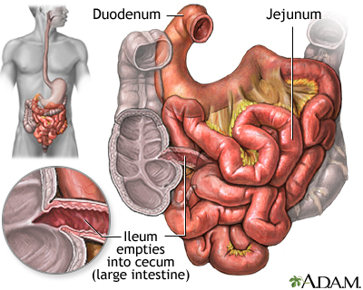
H.3.1 Draw and label a diagram showing a transverse section of the ileum as seen under a light microscope
H.3.2 Explain the structural features of a epithelial cell of a villus as seen in electron micrographs, including microvilli, mitochondria, pinocytotic vesicles and tight junction
H.3.3 Explain the mechanism used by the ileum to absorb and transport food, including facilitated diffusion, active transport and endocytosis
H.3.4 List the materials that are not absorbed and are egested
Option H2: Digestion
H.2.1 State that digestive juices are secreted into the alimentary canal by glands, including salivary glands, gastric glands in the stomach wall, the pancreas and the wall of the small intestine
H.2.2 Explain the structural features of exocrine gland cells
H.2.3 Compare the composition of saliva, gastric juice and pancreatic juice
H.2.4 Outline the control of digestive juice secretion by nerves and hormones, using the example of secretion of gastric juice
H.2.5 Outline the role of membrane-bound enzymes on the surface of epithelial cells in the small intestine in digestion
H.2.6 Outline the reason for cellulose not being digested in the alimentary canal
H.2.7 Explain why pepsin and trypsin are initially synthesized as inactive precursors and how they are subsequently activated
H.2.8 Discuss the roles of gastric acid and Helicobacter pylori in the development of stomach uclers and stomach cancers
H.2.9 Explain the problem of lipid digestion in a hydrophilic medium and the role of bile in overcoming this
Option H1: Hormonal control
H.1.1 State that hormones are chemical messengers secreted endocrine glands into the blood and transported to specific target cells
H.1.2 State that hormones can be steroids, proteins and tyrosine derivatives, with one example of each
H.1.3 Distinguish between the mode of action of steroid hormones and protein hormones
H.1.4 Outline the relationship between the hypothalamus and the pituitary gland
H.1.5 Explain the control of ADH (vasopressin) secretion by negative feedback
2014年7月30日 星期三
Option H: Further human physiology
Option H of the IB HL Biology syllabus is the Further human physiology. IBO recommends to spend 22 hours on this topic.
This topic has 6 sub-chapters: "Hormonal control", "Digestion", "Absorption of digested food", "Function of the liver", "The transport system" and "Gas exchange". Each are separated with numerical values in order of mentioned.
These are all HL option syllabus statements, it is recommended to bring a Casio Graphical Calculator instead of Texas.
2014年6月9日 星期一
Topic 11.4: Reproduction
11.4.1 Annotate a light micrograph of testis tissue to show the location and function of interstitial cells (Leydig cells), germinal epithelium cells, developing spermatozoa and Sertoli cells
11.4.2 Outline the processes involved in spermatogenesis within the testis, including mitosis, cell growth, the two divisions of meiosis and cell differentiation
11.4.3 State the role of LH, testosterone and FSH in spermatogenesis
11.4.4 Annotate a diagram of the ovary to show the location and function of germinal epithelium, primary follicles, mature follicle and secondary oocyte
11.4.5 Outline the processes involved in oogenesis within the ovary, including mitosis, cell growth, the two divisions of meiosis, the unequal division of cytoplasm and the degeneration of polar body
11.4.6 Draw and label a diagram of a mature sperm and egg
11.4.7 Outline the role of the epididymis, seminal vesicle and prostate gland in the production of semen
11.4.8 Compare the processes of spermatogenesis and oogenesis, including the number of gametes and the timing of the formation and release of gametes
11.4.9 Describe the process of fertilization, including the acrosome reaction, penetration of the egg membrane by a sperm and the cortical reaction
11.4.10 Outline the role of HCG in early pregnancy
11.4.11 Outline the early embryo development up to the implantation of the blastocyst
11.4.12 Explain how the structure and functions of the placenta, including its hormonal role in the secretion of estrogen and progesterone, maintain pregnancy
11.4.13 State that the fetus is supported and protected by the amniotic sac and amniotic fluid
11.4.14 State that materials are exchanged between the maternal and fetal blood in the placenta
11.4.15 Outline the process of birth and its hormonal control, including the changes in progesterone and oxytocin levels and positive feedback
Topic 11.3: The kidney
11.3.1 Define excretion
11.3.2 Draw and label a diagram of the kidney
11.3.3 Annotate a diagram of a glomerulus and associated nephron to show the function of each part
11.3.4 Explain the process of ultrafiltration, including blood pressure, fenestrated blood capillaries and basement membrane
11.3.5 Define osmoregulation
11.3.6 Explain the reabsorption of glucose, water and salts in the proximal convulated tubule, including the roles of microvilli, osmosis and active transport
11.3.7 Explain the roles of the loop of Henle, medulla, collecting duct and ADH (vasopressin) in maintaining the water balance of the blood
11.3.8 Explain the differences in the concentration of proteins, glucose and urea between blood plasma, glomerular filtrate and urine
11.3.9 Explain the presence of glucose in the urine of untreated diabetic patients
Topic 11.2: Muscles and movement
11.2.1 State the roles of bones, ligaments, muscles, tendons and nerves in human movement
11.2.2 Label a diagram of the human elbow join, including cartilages, synovial fluid, joint capsule, named bones and antagonistic muscles (biceps and triceps)
11.2.3 Outline the functions of the structures in the human elbow joint named in 11.2.2
11.2.4 Compare the movements in the hip joint and the knee joint
11.2.5 Describe the structure of striated muscle fibres, including the myofibrils with light and dark bands, mitochondria, the sarcoplasmic reticulum, nuclei and the sacrolemma
11.2.6 Draw and label a diagram to show the structure of a sacromere, including Z lines, actin filaments, myosin filaments with heads, and the resultant light and dark bands
11.2.7 Explain how skeletal muscles contracts, including the release of calcium ions from the sacroplasmic reticulum, the formation of cross-bridges, the sliding of actin and myosin filaments, and the use of ATP to break cross-bridges and re-set myosin heads
11.2.8 Analyse electron micrographs to find the state of contraction of muscles fibres
Topic 11.1: Defense against infectious disease
11.1.1 Describe the process of blood clotting
11.1.2 Outline the principle of challenge and response, clonal selection and memory cells as the basis of immunity
11.1.3 Define active and passive immunity
11.1.4 Explain antibody production
11.1.5 Describe the production of monoclonal antibodies and their use in diagnosis and in treatment
11.1.6 Explain the principle of vaccination
11.1.7 Discuss the benefits and dangers of vaccination
Topic 11: Human health and physiology
Topic 11 of the IB HL Biology syllabus is the Genetics. IBO recommends to spend 17 hours on this topic.
This topic has 4 sub-chapters: "Defense against infectious disease", "Muscles and movement", "The kidney" and "Reproduction". Each are separated with numerical values in order of mentioned.
These are all HL syllabus statements, it is recommended to bring a Casio Graphical Calculator instead of Texas.
Topic 6.6: Reproduction
6.6.1 Draw and label diagrams of the adult male and female reproductive systems
6.6.2 Outline the role of hormones in the menstrual cycle, including FSH (follicle stimulating hormones), LH (luteinizing hormone), estrogen and progesterone
6.6.3 Annotate a graph showing hormone levels in the menstrual cycle, illustrating the relationship between changes in hormone level and ovulation, menstruation and thickening of the endometrium
6.6.4 List three roles of testosterone in males
6.6.5 Outline the process of in vitro fertilization (IVF)
6.6.6 Discuss the ethical issues associated with IVF
Topic 6.5: Nerves, hormones and homeostasis
6.5.1 State that the nervous system consists of the central nervous system (CNS) and peripheral nerves, and is composed of cells called neurons that can carry rapid electrical impulses
6.5.2 Draw and label a diagram of the structure of a motor neuron
6.5.3 State that nerve impulses are conducted from recpetors to the CNS by sensory neurons, within the CNS by relay neurons, and from the CNS to effectors by motor neurons
6.5.4 Define resting potential and action potential (depolarization and repolarization)
6.5.5 Explain how a nerve impulse passes along a non-myelinated neuron
6.5.6 Explain the principles of synaptic transmission
6.5.7 State that the endocrine system consists of glands that release hormones that are transported in the blood
6.5.8 State that homeostasis involves maintaining the internal environment between limits, including blood pH, carbon dioxide concentration, blood glucose concentration, body temperature and water balance
6.5.9 Explain that homeostasis involves monitoring levels of variables and correcting changes in levels by negative feedback mechanism
6.5.10 Explain the control of body temperature, including the transfer of heat in blood, and the roles of the hypothalamus, sweat glands, skin arterioles and shivering
6.5.11 Explain the control of blood glucose concentration, including the roles of glucagon, insulin and a and b cells in the pancreatic islets
6.5.12 Distinguish between type I and type II diabetes
Topic 6.4: Gas exchange
6.4.1 Distinguish between ventilation, gas exchange and cell respiration
6.4.2 Explain the need for a ventilation system
6.4.3 Describe the features of alveoli that adapt them to gas exchange
6.4.4 Draw and label a diagram of the ventilation system, including trachea, lungs, bronchi, bronchioles and alveoli
6.4.5 Explain the mechanism of ventilation of the lungs in terms of volume and pressure changes caused by the internal and external intercostal muscles, the diaphragm and abdominal muscles
Topic 6.3: Defense against infectious disease
6.3.1 Define pathogen
A pathogen is an organism or virus that causes a disease. Most, but not all, are microorganisms.

6.3.2 Explain why antibiotics are effective against bacteria but not against viruses
Antibiotics are naturally occuring substances that slow down or kill microorganisms. They are obtained mainly from fungi or bacteria, and are substances which such organisms manufacture in their natural habitats.
Most antibiotics are so extremely effective at disrupting bacterial metabolism that whole populations are quickly suppressed. A key process here is the formation and laying down of the new dividing wall. Bacterial cells have a rigid wall containing giant, complex, molecules of amino-sugars and peptide units (polymers) rather than the polysaccharides of plant cell walls.

Virus on the other hand, are not living cells and have no metabolism of their own to be interfered with. Viruses reproduce using metabolic pathways in their host cell that are not affected by antibiotics. Antibiotics cannot be used to prevent viral diseases.

6.3.3 Outline the role of skin and mucous membranes in defense against pathogens
6.3.4 Outline how phagocytes leucocytes ingest pathogens in the blood and in blood tissues
6.3.5 Distinguish between antigens and antibodies
6.3.6 Explain antibody production
6.3.7 Outline the effects of HIV on the immune system
6.3.8 Discuss the cause, transmission and social implications of AIDs
A pathogen is an organism or virus that causes a disease. Most, but not all, are microorganisms.
6.3.2 Explain why antibiotics are effective against bacteria but not against viruses
Antibiotics are naturally occuring substances that slow down or kill microorganisms. They are obtained mainly from fungi or bacteria, and are substances which such organisms manufacture in their natural habitats.
Most antibiotics are so extremely effective at disrupting bacterial metabolism that whole populations are quickly suppressed. A key process here is the formation and laying down of the new dividing wall. Bacterial cells have a rigid wall containing giant, complex, molecules of amino-sugars and peptide units (polymers) rather than the polysaccharides of plant cell walls.
Virus on the other hand, are not living cells and have no metabolism of their own to be interfered with. Viruses reproduce using metabolic pathways in their host cell that are not affected by antibiotics. Antibiotics cannot be used to prevent viral diseases.
6.3.3 Outline the role of skin and mucous membranes in defense against pathogens
6.3.4 Outline how phagocytes leucocytes ingest pathogens in the blood and in blood tissues
6.3.5 Distinguish between antigens and antibodies
6.3.6 Explain antibody production
6.3.7 Outline the effects of HIV on the immune system
6.3.8 Discuss the cause, transmission and social implications of AIDs
Topic 6.2: The transport system
6.2.1 Draw and label a diagram of the heart showing the four chambers, associated blood vessels, valves and the route of blood through the heart

6.2.2 State that the coronary arteries supply heart muscle with oxygen and nutrients
The heart is a muscle itself, so note that the walls of the heart are supplied with oxygenated blood via coronary arteries. These arteries and the capillaries they serve, deliver to the muscle fibres of the heart the oxygen and the nutrients essential for the pumping action.

6.2.3 Explain the action of the heart in terms of collecting blood, pumping blood, and opening and closing of valves
The cavity of the heart is divided into four chambers, with those on the right side of the heart completely separate from those on the left. The two upper chambers are called the artia and these receive blood into the heart.
The lower chambers are thick-walled ventricles, with the muscular wall of the left ventricle much thicker than that of the right ventricle. However, the volumes of the right and left sides (the quantities of blood they contain) are identical. The ventricles pump blood out of the heart.
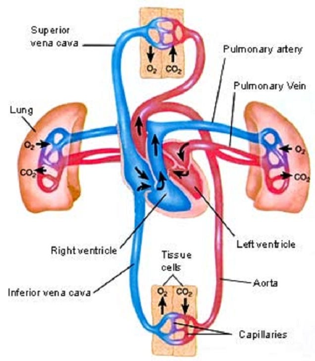
The valves of the heart prevent backflow of the blood, thereby maintaining the direction of flow through the heart. The artio-ventricular valves are large valves, positioned to prevent backflow from ventricles to artia. The edges of these valves are supported by tendons anchored to the muscle walls of the ventricles below. A different type of valve separates the ventricles from the pulmonary artery and the aorta. These are called the semi-lunar valves.

6.2.4 Outline the control of the heartbeat in terms of myogenic muscle contraction, the role of the pacemaker, nerves, the medulla of the brain and epinephrine (adrenaline)
The heart normally beats about 75 times per minute - approximately 0.8 seconds per beat. In each beat, the heart muscle contracts strongly, followed by a period of relaxation.
The atrium contacts pushing blood past the bicuspid valve into the ventricle, Atrium muscle relax. Ventricle muscles contract, causing blood pressure to close the bicuspid valve and open the semilunar valves, forcing blood into the aorta. Ventricle and atrium muscle relax, while the pressure of blood in the aorta causes the semilunar valves to shut. Blood flows into the atrium and opens the bicuspid valve as it starts to flow into the ventricle.
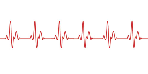 The heart beats rhythmically throughout life, without rest, apart from the momentary relaxation between beats. Even more remarkably, the origin of each beat is within the heart itself - we say the heart beat is myogenic in origin.
Beats originate in a strucuture in the muscle of the wall of the right atrium, called the pacemaker. Special muscle fibre radiate out from the pacemaker, conducting the impulse to the muscles of both artia, triggering contraction. Then a second excitation is passed onto the ventricles causing a ventricular contraction.

The heart receives impulses from a control centre in the hindbrain (medulla), via two nerves. One nerve, when stimulated, triggers speeding up of the heart rate, and the other nerve triggers a slowing down of the heart. Since these nerves have opposite effect, they are antagonistic.

The hormone adrenaline is secreted by the adrenal glands and carried in the blood, causes the pacemaker to increase the heart rate.
6.2.5 Explain the relationship between the structure and function of arteries, capillaries and veins
There are three types of vessels in the circulation system.
Arteries
- Carry blood away from the heart
- Carry blood at high pressures
- Have a narrow lumen
- Have a thick middle layer to help the pulse flow
- Have a outer layer to prevent rupture
Capillaries
- Are a fine networks of tiny tubes linking arteries and veins.
- Involved with material and gas exchange with the surrounding body tissues
- Blood pressure is relatively low
- Small diametre
- Single cell to allow easier diffusion
- Contains pores to aid the transport of materials
Veins
- Carry blood to the heart
- Carry blood at low pressure
- Very wide lumen
- Have valves to prevent blooding pooling
- Thin surrounding tissue as blood do not go in rhythmic pulse.

6.2.6 State that blood is composed of plasma, erythrocytes, leucocytes (phagocytes and lymphocytes) and platelets
Blood is a special tissue consisting of a liquid medium called plasma in which are suspended red cells or erythrocytes, white cells or luecocytes, and platelets. The plasma is the medium for exchange of substances between cells and tissues, the erythrocytes are involved in transport of respiratory gases.

6.2.7 State that the following are transported by the blood: nutrients, oxygen, carbon dioxide, hormones, antibodies, urea and heat
The blood circulation has roles in the body's defense against diseases as well as being the all-important transport system of the body. Nutrients from digestion, oxygen and carbon dioxide, urea, hormones and antibodies are all transported. In the tissues of the body, exchange between the blood and cells of the tissues occurs from the capillaries, the walls of which are permeable and highly "leaky".
Tissue respiration - gas exchange
Hydration - water to all the tissues
Nutrition - Nutrients (sugars, amino acids, lipids, vitamins) and inorganic ions to all cells.
Excretion - Waste product urea to kidneys
Development and co-ordination - Hormones from endocrine glands to target organs
Temperature regulation - distribution of heat
Defense against disease - Antibodies are circulated in the blood system

6.2.2 State that the coronary arteries supply heart muscle with oxygen and nutrients
The heart is a muscle itself, so note that the walls of the heart are supplied with oxygenated blood via coronary arteries. These arteries and the capillaries they serve, deliver to the muscle fibres of the heart the oxygen and the nutrients essential for the pumping action.
6.2.3 Explain the action of the heart in terms of collecting blood, pumping blood, and opening and closing of valves
The cavity of the heart is divided into four chambers, with those on the right side of the heart completely separate from those on the left. The two upper chambers are called the artia and these receive blood into the heart.
The lower chambers are thick-walled ventricles, with the muscular wall of the left ventricle much thicker than that of the right ventricle. However, the volumes of the right and left sides (the quantities of blood they contain) are identical. The ventricles pump blood out of the heart.
The valves of the heart prevent backflow of the blood, thereby maintaining the direction of flow through the heart. The artio-ventricular valves are large valves, positioned to prevent backflow from ventricles to artia. The edges of these valves are supported by tendons anchored to the muscle walls of the ventricles below. A different type of valve separates the ventricles from the pulmonary artery and the aorta. These are called the semi-lunar valves.
6.2.4 Outline the control of the heartbeat in terms of myogenic muscle contraction, the role of the pacemaker, nerves, the medulla of the brain and epinephrine (adrenaline)
The heart normally beats about 75 times per minute - approximately 0.8 seconds per beat. In each beat, the heart muscle contracts strongly, followed by a period of relaxation.
The atrium contacts pushing blood past the bicuspid valve into the ventricle, Atrium muscle relax. Ventricle muscles contract, causing blood pressure to close the bicuspid valve and open the semilunar valves, forcing blood into the aorta. Ventricle and atrium muscle relax, while the pressure of blood in the aorta causes the semilunar valves to shut. Blood flows into the atrium and opens the bicuspid valve as it starts to flow into the ventricle.
Beats originate in a strucuture in the muscle of the wall of the right atrium, called the pacemaker. Special muscle fibre radiate out from the pacemaker, conducting the impulse to the muscles of both artia, triggering contraction. Then a second excitation is passed onto the ventricles causing a ventricular contraction.
The heart receives impulses from a control centre in the hindbrain (medulla), via two nerves. One nerve, when stimulated, triggers speeding up of the heart rate, and the other nerve triggers a slowing down of the heart. Since these nerves have opposite effect, they are antagonistic.
The hormone adrenaline is secreted by the adrenal glands and carried in the blood, causes the pacemaker to increase the heart rate.
6.2.5 Explain the relationship between the structure and function of arteries, capillaries and veins
There are three types of vessels in the circulation system.
Arteries
- Carry blood away from the heart
- Carry blood at high pressures
- Have a narrow lumen
- Have a thick middle layer to help the pulse flow
- Have a outer layer to prevent rupture
Capillaries
- Are a fine networks of tiny tubes linking arteries and veins.
- Involved with material and gas exchange with the surrounding body tissues
- Blood pressure is relatively low
- Small diametre
- Single cell to allow easier diffusion
- Contains pores to aid the transport of materials
Veins
- Carry blood to the heart
- Carry blood at low pressure
- Very wide lumen
- Have valves to prevent blooding pooling
- Thin surrounding tissue as blood do not go in rhythmic pulse.
6.2.6 State that blood is composed of plasma, erythrocytes, leucocytes (phagocytes and lymphocytes) and platelets
Blood is a special tissue consisting of a liquid medium called plasma in which are suspended red cells or erythrocytes, white cells or luecocytes, and platelets. The plasma is the medium for exchange of substances between cells and tissues, the erythrocytes are involved in transport of respiratory gases.
6.2.7 State that the following are transported by the blood: nutrients, oxygen, carbon dioxide, hormones, antibodies, urea and heat
The blood circulation has roles in the body's defense against diseases as well as being the all-important transport system of the body. Nutrients from digestion, oxygen and carbon dioxide, urea, hormones and antibodies are all transported. In the tissues of the body, exchange between the blood and cells of the tissues occurs from the capillaries, the walls of which are permeable and highly "leaky".
Tissue respiration - gas exchange
Hydration - water to all the tissues
Nutrition - Nutrients (sugars, amino acids, lipids, vitamins) and inorganic ions to all cells.
Excretion - Waste product urea to kidneys
Development and co-ordination - Hormones from endocrine glands to target organs
Temperature regulation - distribution of heat
Defense against disease - Antibodies are circulated in the blood system
Topic 6.1: Digestion
6.1.1 Explain why digestion of large food molecules is essential.
Most food is solid and in the form of large complex molecules which are insoluble and chemically inert (not readily stable)
Large molecules need to be broken down into smaller molecules that can be readily absorbed across membranes and into cells. Small molecules can be reassembled into new products (e.g. amino acids can be reassembled to form new proteins).

6.1.2 Explain the need for enzymes in digestion
Enzymes are biological catalysts that speeds up the rate of reaction. It allows reactions to take place at body temperatures. They are specific for a certain type of reaction though based on the lock and key model.

6.1.3 State the source, substrate, products and optimum pH conditions for one amylase, one protease and one lipase
Amylase
Example - Salivary amylase
Source - Salivary glands
Substrate - Starch
Product - Maltose
Optimum pH - 7
Protease
Example - Pepsin
Source - Gastric juice
Substrate - Protein
Product - Short polypeptides
Optimum pH - 2
Lipase
Example - Pancreatic juice
Source - Pancreas
Substrate - Lipids (triglyceride)
Product - Glycerol and fatty acids
Optimum pH - 8
6.1.4 Draw and label a diagram of the digestive system

6.1.5 Outline the function of the stomach, small intestine and large intestine
Stomach
Present in the wall of the stomach are millions of tiny pits called gastric glands which secrete the components of gastric juice. This juice includes hydrochloric acid - sufficiently acidic to create an environment of pH 1.5-2.0, which is the optimum pH for protein digestion by the protease enzymes of the gastric juice. These proteases, of which pepsi is one, are formed in cells of gastric glands and secreted in an inactive state. The hydrochloric acid then activates them, and kills most of the in coming bacteria in the food.
The whole stomach lining is supplied with goblet cells that secrete mucus. Mucus bathes the interior lining of the stomach, forming an effective barrier to both the hydrochloric acid and the protease of the gastric juices, preventing autolysis (self-digestion) of the stomach wall.
As the food is mixed with gastric juice and churned by muscle action it becomes a semi-liquid called chyme. The churning action of the stomach is an important part of the mechanical digestion process. A typical meal may spend up to four hours in the stomach.

Small intestines
Food enters the first part of the small intestine (known as the duodenum) a little at a time. Here the chyme meets bile from the bile duct, and the pancreatic juice from the pancreas. Bile is strongly alkaline and neutralises the acidity of the chyme. It also lowers the surface tension of large fat globules, causing them to break into tiny droplets, a process called emulsification. This speeds digestion by the enzyme lipase later on. Bile itself contains no enzyme.
All these enzymes act as the chyme, bile and pancreatic juice are mixed together by a churning action (a form of peristalsis) called segmentation.
6.1.6 Distinguish between absorption and assimilation
Absorption can be defined as the movement of particles or dissolved substances across a membrane
Assimilation can be defined as the conversion of nutrients into fluids or solid parts of an organism.

6.1.7 Explain how the structure of the villus is related to its role in absorption and transport of the products of digestion
There are different structure of the villus which aid its role in absorption.
Micro-villi is present to provide a huge surface area for absorption.
Epithelium cells has a single layer of small cells, packed with mitochondria - the source of ATP for active uptake across the plasma membrane.
Protein pumps in the plasma membrane of the epithelial cells can actively transport nutrients across the plasma membrane into the villi
There is also a big network of capillaries which proved a large surface area for uptake of amino acids, monosaccharides, and fatty acids and glycerol into blood circulation.
Lacteal is a branch of the lymphatic system into which triglycerides (combined with protein) pass for transport to body cells
The mucus from goblet cells in the epithelium can lubricate movement of digested food among the villi and protects plasma membrane of epithelial cells.

Most food is solid and in the form of large complex molecules which are insoluble and chemically inert (not readily stable)
Large molecules need to be broken down into smaller molecules that can be readily absorbed across membranes and into cells. Small molecules can be reassembled into new products (e.g. amino acids can be reassembled to form new proteins).
6.1.2 Explain the need for enzymes in digestion
Enzymes are biological catalysts that speeds up the rate of reaction. It allows reactions to take place at body temperatures. They are specific for a certain type of reaction though based on the lock and key model.
6.1.3 State the source, substrate, products and optimum pH conditions for one amylase, one protease and one lipase
Amylase
Example - Salivary amylase
Source - Salivary glands
Substrate - Starch
Product - Maltose
Optimum pH - 7
Protease
Example - Pepsin
Source - Gastric juice
Substrate - Protein
Product - Short polypeptides
Optimum pH - 2
Lipase
Example - Pancreatic juice
Source - Pancreas
Substrate - Lipids (triglyceride)
Product - Glycerol and fatty acids
Optimum pH - 8
6.1.4 Draw and label a diagram of the digestive system
6.1.5 Outline the function of the stomach, small intestine and large intestine
Stomach
Present in the wall of the stomach are millions of tiny pits called gastric glands which secrete the components of gastric juice. This juice includes hydrochloric acid - sufficiently acidic to create an environment of pH 1.5-2.0, which is the optimum pH for protein digestion by the protease enzymes of the gastric juice. These proteases, of which pepsi is one, are formed in cells of gastric glands and secreted in an inactive state. The hydrochloric acid then activates them, and kills most of the in coming bacteria in the food.
The whole stomach lining is supplied with goblet cells that secrete mucus. Mucus bathes the interior lining of the stomach, forming an effective barrier to both the hydrochloric acid and the protease of the gastric juices, preventing autolysis (self-digestion) of the stomach wall.
As the food is mixed with gastric juice and churned by muscle action it becomes a semi-liquid called chyme. The churning action of the stomach is an important part of the mechanical digestion process. A typical meal may spend up to four hours in the stomach.
Small intestines
Food enters the first part of the small intestine (known as the duodenum) a little at a time. Here the chyme meets bile from the bile duct, and the pancreatic juice from the pancreas. Bile is strongly alkaline and neutralises the acidity of the chyme. It also lowers the surface tension of large fat globules, causing them to break into tiny droplets, a process called emulsification. This speeds digestion by the enzyme lipase later on. Bile itself contains no enzyme.
All these enzymes act as the chyme, bile and pancreatic juice are mixed together by a churning action (a form of peristalsis) called segmentation.
6.1.6 Distinguish between absorption and assimilation
Absorption can be defined as the movement of particles or dissolved substances across a membrane
Assimilation can be defined as the conversion of nutrients into fluids or solid parts of an organism.
6.1.7 Explain how the structure of the villus is related to its role in absorption and transport of the products of digestion
There are different structure of the villus which aid its role in absorption.
Micro-villi is present to provide a huge surface area for absorption.
Epithelium cells has a single layer of small cells, packed with mitochondria - the source of ATP for active uptake across the plasma membrane.
Protein pumps in the plasma membrane of the epithelial cells can actively transport nutrients across the plasma membrane into the villi
There is also a big network of capillaries which proved a large surface area for uptake of amino acids, monosaccharides, and fatty acids and glycerol into blood circulation.
Lacteal is a branch of the lymphatic system into which triglycerides (combined with protein) pass for transport to body cells
The mucus from goblet cells in the epithelium can lubricate movement of digested food among the villi and protects plasma membrane of epithelial cells.
Topic 6: Human health and physiology
Topic 6 of the IB SL Biology syllabus is the Human health and physiology. IBO recommends to spend 20 hours on this topic.
This topic has 6 sub-chapters: "Digestion", "The transport system", "Defense against infectious defense", "Gas exchange", "Nerves, hormones and homeostasis" and "Reproduction". Each are separated with numerical values in order of mentioned.
These are all SL syllabus statements, it is recommended to bring a Casio Graphical Calculator instead of Texas.
2014年4月13日 星期日
Topic 10.3: Polygenic inheritance
10.3.1 Define polygenic inheritance
Polygenic inheritance refers to a single characteristic that is controlled by more than two genes (also called multifactorial inheritance)
Polygenic inheritance patterns normally (bell-shaped) distribution curve - it shows continuous variation
By increasing the number of genes controlling a trait, the number of phenotype combinations also increase, until the number of phenotypes to which an individual can be assigned are no longer discrete, but continuous

10.3.2 Explain that polygenic inheritance can contribute to continuous variation using two examples, one of which must be human skin colour
Human skin colour

Polygenic inheritance refers to a single characteristic that is controlled by more than two genes (also called multifactorial inheritance)
Polygenic inheritance patterns normally (bell-shaped) distribution curve - it shows continuous variation
By increasing the number of genes controlling a trait, the number of phenotype combinations also increase, until the number of phenotypes to which an individual can be assigned are no longer discrete, but continuous
10.3.2 Explain that polygenic inheritance can contribute to continuous variation using two examples, one of which must be human skin colour
Human skin colour
- The colour of human skin is determined by the amount of dark pigment (melanin) it contains
- At least four (possibly more) genes are involved in melanin production; for each gene one allele codes for melanin production, the other does not
- The combination of melanin producing alleles determines the degree of pigmentation, leading to continuous variation
Grain colour in wheat
- Wheat grains vary in colour from white to dark red, depending on the amount of red pigment they contain
- Three genes control the colour and each gene has two alleles (one coding for red pigment, the other coding for no pigment)
- The most frequent combinations have an equal number of "pigment producing" and "no pigment" alleles, whereas combinations of one extreme or the other are relatively rare
- The overall pattern of inheritance shows continuous variation
Topic 10.2: Dihybrid crosses and gene linkage
10.2.1 Calculate and predict the genotypic and phenotypic ratio of offspring of dihybrid crosses involving unlinked autosomal genes
A dihybrid cross determines the allele combinations of offspring for two particular genes that are unlinked (not on the same chromosomes)
Because there are two genes with two alleles per gene (multiple alleles not required), there can be up to four different gamete combinations

10.2.2 Distinguish between autosomes and sex chromosomes
Autosomes: Pairs of chromosomes that are identical in appearance (e.g. same size, same gene loci, etc) and are not involved in sex determination
Sex chromosome: Pairs of chromosomes involved in sex determination and are not identical in appearance (e.g. X and Y chromosome in humans)

10.2.3 Explain how crossing over between non-sister chromatids of a homologous pair in prophase I can result in an exchange in alleles
During crossing over in prophase I, non-sister chromatids of a homologous pair may break and reform at points of attachment called chiasmata
As these chromatids break at the same point, any gene loci below the point of the break will be exchanged as a result of recombination
This means that maternal and paternal alleles may be exchanged between the maternal and paternal chromosomes, creating new gene combinations
The further apart two gene loci are on a chromosome, the more likely they are to be exchanged

10.2.4 Define linkage group
A linkage group is a group of genes whose loci are on the same chromosome and therefore do not follow the law of independent assortment
Linkage genes will tend to be inherited together - the only way to separate them is through recombination (via crossing over during synapsis)
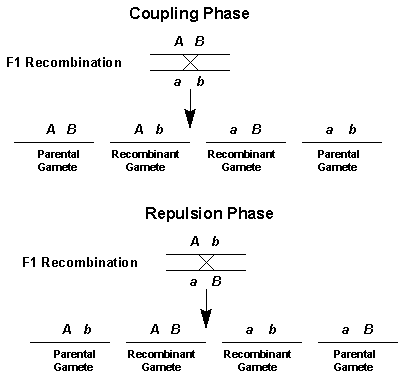
10.2.5 Explain an example of a cross linkage between two linked genes
When two genes are linked, they do not follow the expected phenotypic ratio for a dihybrid cross between heterozygous parents
Instead the phenotypic ratio will follow that of a monohybrid cross as the two genes are inherited together
This means that offspring will tend to produce the parental phenotypes
Recombinant phenotypes will only be evident if crossing over occurs in prophase I and would thus be expected to appear in low number (if at all)
An example of a cross between two linked genes is the mating of a grey bodied, normal wing fruit fly with a black bodied, vestigial wing mutant

10.2.6 Identify which of the offspring are recombinants in a dihybrid cross involving linked genes
Recombinants of linked genes are those combinations of genes not found in parents
For example, in a test cross of a heterozygous fruit fly (grey bodied, normal wings) with a homozygous recessive mutant (black bodied, vestigial wings), the recombinants would be the grey bodied, vestigial winged offsprings and the black bodied, normal winged offspring

Linked genes that have undergone recombination can be distinguished from unlinked genes via test cross because the frequency of the recombinant genotype will always be less than would occur for unlinked genes (crossing over does not happen every time)
A dihybrid cross determines the allele combinations of offspring for two particular genes that are unlinked (not on the same chromosomes)
Because there are two genes with two alleles per gene (multiple alleles not required), there can be up to four different gamete combinations
10.2.2 Distinguish between autosomes and sex chromosomes
Autosomes: Pairs of chromosomes that are identical in appearance (e.g. same size, same gene loci, etc) and are not involved in sex determination
Sex chromosome: Pairs of chromosomes involved in sex determination and are not identical in appearance (e.g. X and Y chromosome in humans)
10.2.3 Explain how crossing over between non-sister chromatids of a homologous pair in prophase I can result in an exchange in alleles
During crossing over in prophase I, non-sister chromatids of a homologous pair may break and reform at points of attachment called chiasmata
As these chromatids break at the same point, any gene loci below the point of the break will be exchanged as a result of recombination
This means that maternal and paternal alleles may be exchanged between the maternal and paternal chromosomes, creating new gene combinations
The further apart two gene loci are on a chromosome, the more likely they are to be exchanged
10.2.4 Define linkage group
A linkage group is a group of genes whose loci are on the same chromosome and therefore do not follow the law of independent assortment
Linkage genes will tend to be inherited together - the only way to separate them is through recombination (via crossing over during synapsis)
10.2.5 Explain an example of a cross linkage between two linked genes
When two genes are linked, they do not follow the expected phenotypic ratio for a dihybrid cross between heterozygous parents
Instead the phenotypic ratio will follow that of a monohybrid cross as the two genes are inherited together
This means that offspring will tend to produce the parental phenotypes
Recombinant phenotypes will only be evident if crossing over occurs in prophase I and would thus be expected to appear in low number (if at all)
An example of a cross between two linked genes is the mating of a grey bodied, normal wing fruit fly with a black bodied, vestigial wing mutant
10.2.6 Identify which of the offspring are recombinants in a dihybrid cross involving linked genes
Recombinants of linked genes are those combinations of genes not found in parents
For example, in a test cross of a heterozygous fruit fly (grey bodied, normal wings) with a homozygous recessive mutant (black bodied, vestigial wings), the recombinants would be the grey bodied, vestigial winged offsprings and the black bodied, normal winged offspring
Linked genes that have undergone recombination can be distinguished from unlinked genes via test cross because the frequency of the recombinant genotype will always be less than would occur for unlinked genes (crossing over does not happen every time)
- For example:
- Heterozygous test cross of unlinked genes = 1:1:1:1 phenotypic ratio
- Heterozygous test cross of linked genes = 1:1:0.1:0.1 pehnotypic ratio (uncommon phenotypes are recombinants)
Topic 10.1: Meiosis
10.1.1 Describe the behaviour of the chromosomes in the phases of meiosis
Interphase: Cell growth and DNA replication (duplication of DNA creates sister chromatid chromosome)
Meiosis I
10.1.2 Outline the formation of chiasmata in the process of crossing over
Crossing over involves the exchange of segments of DNA between homologous chromosomes during Prophase I of meiosis
The process of crossing over occurs as follows

10.1.3 Explain how meiosis results in an effectively infinite genetic variety in gametes through crossing over in prophase I and random orientation in metaphase I
During anaphase I, homologous chromosomes separate, such that each resultant daughter (and subsequent gametes) contains a chromosome of either maternal or paternal origin
The orientation of these homologous in metaphase I is random, such that there is an equal probability of the daughter cell having either the maternal or paternal chromosome
As humans have a haploid number of 23 chromosomes, this means that there is 223 potential gamete combinations (over 8 million combinations)
Crossing over in prophase I results in entirely new chromosome combinations, as recombination through gene exchange produces wholly original chromosomes containing both maternal and paternal DNA, resulting in near infinite genetic variability
Other sources of genetic variation include random fertilisations, DNA mutations, chromosome mutations and non-disjunction

10.1.4 State Mendal's law of independent assortment
Gregor Mendal was a 19th century Moravian monk who demonstrated that the inheritance of traits (i.e. genes) followed particular laws:

10.1.5 Explain the relationship between Mendal's law of independent assortment and meiosis
The law of independent assortment relates to the random orientation of homologous chromosomes in metaphase I of meiosis
Because the orientation of a homologous pair is random, and does not affect the orientation of any other homologous pair, any one of a pair of alleles on a chromosome has an equal chance of being paired with, or separated from, any one of a pair of alleles on another chromosome
This means the inheritance of two different traits will occur independently of each other (provided the genes aren't linked)

Interphase: Cell growth and DNA replication (duplication of DNA creates sister chromatid chromosome)
Meiosis I
- Prophase I: DNA supercoils and chromosomes condense, nuclear membrane dissolves, homologous pairs form bivalents, crossing over occurs
- Metaphase I: Spindle fibres from centrioles (at poles) attach to centromeres of bivalent, bivalents line up along the equator of the cell
- Anaphase I: Spindle fibres contract and split the bivalent, homologous chromosomes move to opposite poles of the cell
- Telophase I: Chromosomes decondense, nuclear membranes may reform, cell divide (cytokinesis) forming two haploid daughters cells
Interkinesis: An optional rest period between meiosis I and meiosis II, no DNA replication occurs in this stage
Meiosis II

- Prophase II: Chromosomes condense, nuclear membrane dissolves (if reformed), centrioles move to opposite poles (perpendicular to previous poles)
- Metaphase II: Spindle fibres form centrioles attach to centromeres of chromosomes, chromosomes line up along the equator of the cell
- Anaphase II: Spindle fibres contract and split the chromosome into sister chromatids, chromatids (now called chromosomes) move to opposite poles
- Telophase II: Chromosomes decondense, nuclear membrane reforms, cells divide (cytokinesis), resulting in four haploid daughter cells
10.1.2 Outline the formation of chiasmata in the process of crossing over
Crossing over involves the exchange of segments of DNA between homologous chromosomes during Prophase I of meiosis
The process of crossing over occurs as follows
- Homologous chromosomes become connected in a process called synapsis, forming a bivalent (or tetrad)
- Non-sister chromatids break and recombine with their homologous partner, effectively exchanging genetic material (crossing over)
- The non-sister chromatids remain connected in an X-shaped structure and the positions of attachment are called chiasmata
Chiasmata hold homologous chromosomes together as a bivalent until anaphase I
As a result of crossing over, chromatids may consist of a combination of DNA derived from both homologous - these are called recombinant
10.1.3 Explain how meiosis results in an effectively infinite genetic variety in gametes through crossing over in prophase I and random orientation in metaphase I
During anaphase I, homologous chromosomes separate, such that each resultant daughter (and subsequent gametes) contains a chromosome of either maternal or paternal origin
The orientation of these homologous in metaphase I is random, such that there is an equal probability of the daughter cell having either the maternal or paternal chromosome
As humans have a haploid number of 23 chromosomes, this means that there is 223 potential gamete combinations (over 8 million combinations)
Crossing over in prophase I results in entirely new chromosome combinations, as recombination through gene exchange produces wholly original chromosomes containing both maternal and paternal DNA, resulting in near infinite genetic variability
Other sources of genetic variation include random fertilisations, DNA mutations, chromosome mutations and non-disjunction
10.1.4 State Mendal's law of independent assortment
Gregor Mendal was a 19th century Moravian monk who demonstrated that the inheritance of traits (i.e. genes) followed particular laws:
- Law of segregation: Each herediatary characteristics is controlled by two alleles, which segregate and pass into different reproductive cells (gametes)
- Law of independent assortment: The separation of alleles for one gene will occur independently of the separation of alleles for another gene
- According to the law of independent assortment, different allele combinations should always be equally impossible
- However this law only holds for genes that are on different chromosomes - the law of independent assortment does not apply to linked genes
10.1.5 Explain the relationship between Mendal's law of independent assortment and meiosis
The law of independent assortment relates to the random orientation of homologous chromosomes in metaphase I of meiosis
Because the orientation of a homologous pair is random, and does not affect the orientation of any other homologous pair, any one of a pair of alleles on a chromosome has an equal chance of being paired with, or separated from, any one of a pair of alleles on another chromosome
This means the inheritance of two different traits will occur independently of each other (provided the genes aren't linked)
Topic 10: Genetics
Topic 10 of the IB HL Biology syllabus is the Genetics. IBO recommends to spend 6 hours on this topic.
This topic has 3 sub-chapters: "Meiosis", "Dihybrid crosses and gene linkage" and "Polygenic inheritance". Each are separated with numerical values in order of mentioned.
These are all HL syllabus statements, it is recommended to bring a Casio Graphical Calculator instead of Texas.
This topic has 3 sub-chapters: "Meiosis", "Dihybrid crosses and gene linkage" and "Polygenic inheritance". Each are separated with numerical values in order of mentioned.
These are all HL syllabus statements, it is recommended to bring a Casio Graphical Calculator instead of Texas.
2014年3月28日 星期五
Topic 4.4: Genetic engineering and biotechnology
4.4.1 Outline the use of polymerase chain reaction (PCR) to copy and amplify minute quantities of DNA
PCR is a way of producing large quantities of a specific target sequences of DNA
It is useful when only a small amount of DNA is available for testing
PCR occurs in a thermal cycler and involves a repeat procedure of 3 steps:

4.4.2 State that, in gel electrophoresis, fragments of DNA move in an electric field and are separated according to their size.
Gel electrophoresis is a technique which is used to separate fragments of DNA according in size

4.4.3 State that get electrophoresis of DNA is used in DNA profiling.
DNA profiling is a technique by which individuals are identified on the basis of their respective DNA profiles
Within the non-coding region of an individual's genome, there exists satellite DNA - long stretches of DNA made up of repeating elements called short tandem repeats (STRs)
These repeating sequences can be existed to form fragments, by cutting with a variety of restriction endonucleases (which cut DNA at specific sites)
As individuals all have a different number of repeats in a given sequence of satellite DNA, they will all generate unique fragment profiles
These different profiles can be compared using gel electrophoresis

4.4.4 Describe the application of DNA profiling to determine paternity and also in forensic investigations.
A DNA sample is collected (blood, saliva, semen, etc) and amplied using PCR
Satellite DNA (non-coding) is cut with specific restriction enzymes to generate fragments
Individuals will have unique fragment lengths due to the variable length of their short tandem repeats (STR)
The fragments are separated with gel electrophoresis (smaller fragments move quickers through the gel)
The DNA profile can then be analysed according to need
Two applications of DNA profiling are:

4.4.5 Analyse DNA profiles to draw conclusions about paternity or forensic investigations
Paternity testing: Children inherit half of their alleles from each parent and thus should possess a combination of their parents alleles
Forensic investigation: Suspect DNA should be a complete match with the sample taken from a crime scene if a conviction is to occur

4.4.6 Outline three outcomes of the sequencing of the complete human genome
The human genome project (HGP) was an international cooperative venture established to sequence the 3 billion base pair (~25,000 genes) in the human genome
The outcomes of this project include:

4.4.7 State that, when genes are transferred between species, the amino acid sequence of polypeptides translated from them is unchanged because the genetic code is universal.
The genetic code is universal, meaning that for every living organism the same codons code for the same amino acids (there are few rare exceptions)
This means that the genetic information from one organism could be translated by another (i.e. it is theoretically transferable)
4.4.8 Outline a basic technique used for gene transfer involving plasmids, a host cell (bacterium, yeast or other cell), restriction enzymes (endonucleases) and DNA ligase
1. DNA Extraction

4.4.9 State two examples of the current uses of genetically modified crops or animals
Crops

4.4.10 Discuss the potential benefits and possible harmful effects of one example of genetic modification
Potential benefits

4.4.11 Define clone
A clone is a group of genetically identical organisms or a group cells derived from a single parent cells

4.4.12 Outline a technique for cloning using differentiated animal cells
Somatic cell nucleus transfer (SCNT) is a method of reproductive cloning using differentiated animal cells

4.4.13 Discuss the ethical issues of therapeutic cloning in humans.
Arguments for therapuetic cloning

PCR is a way of producing large quantities of a specific target sequences of DNA
It is useful when only a small amount of DNA is available for testing
PCR occurs in a thermal cycler and involves a repeat procedure of 3 steps:
- Denaturation: DNA sample is heated to separate it into two strands
- Annealing: DNA primers attached to opposite ends of the target sequences
- Elongation: A heat-tolerant DNA polymerase (Taq) copies the strand
One cycle of PCR yields two identical copies the DNA sequences
- A standard reaction of 30 cycles would yield 1,073,741,826 copies of DNA
4.4.2 State that, in gel electrophoresis, fragments of DNA move in an electric field and are separated according to their size.
Gel electrophoresis is a technique which is used to separate fragments of DNA according in size
- Samples of fragmented DNA are placed in the wells of an agarose gel
- The gel is placed in a buffering solution and an electrical current is passed across the gel
- DNA, being negatively charged (due to phosphate), moves to the positive terminus (anode)
- Smaller fragments are less impeded by the gel matrix and move faster through the gel
- The fragments are thus separated according to size
- Size can be calculated (in kilobases) by comparing against a known industry standard
4.4.3 State that get electrophoresis of DNA is used in DNA profiling.
DNA profiling is a technique by which individuals are identified on the basis of their respective DNA profiles
Within the non-coding region of an individual's genome, there exists satellite DNA - long stretches of DNA made up of repeating elements called short tandem repeats (STRs)
These repeating sequences can be existed to form fragments, by cutting with a variety of restriction endonucleases (which cut DNA at specific sites)
As individuals all have a different number of repeats in a given sequence of satellite DNA, they will all generate unique fragment profiles
These different profiles can be compared using gel electrophoresis
4.4.4 Describe the application of DNA profiling to determine paternity and also in forensic investigations.
A DNA sample is collected (blood, saliva, semen, etc) and amplied using PCR
Satellite DNA (non-coding) is cut with specific restriction enzymes to generate fragments
Individuals will have unique fragment lengths due to the variable length of their short tandem repeats (STR)
The fragments are separated with gel electrophoresis (smaller fragments move quickers through the gel)
The DNA profile can then be analysed according to need
Two applications of DNA profiling are:
- Paternity testing (comparing DNA of offspring against potential fathers)
- Forensic investigation (identifying suspects or victims based on crime-scene DNA)
4.4.5 Analyse DNA profiles to draw conclusions about paternity or forensic investigations
Paternity testing: Children inherit half of their alleles from each parent and thus should possess a combination of their parents alleles
Forensic investigation: Suspect DNA should be a complete match with the sample taken from a crime scene if a conviction is to occur
4.4.6 Outline three outcomes of the sequencing of the complete human genome
The human genome project (HGP) was an international cooperative venture established to sequence the 3 billion base pair (~25,000 genes) in the human genome
The outcomes of this project include:
- Mapping: We now know the number, location and basic sequence of human genes
- Screening: This has allowed for the production of specific gene probes to detect sufferers and carriers of genetic disease conditions
- Medicine: With the discovery of new proteins and their functions, we can develop improved treatments (pharmacogenetics and rational drug design)
- Ancestry: It will give us improved insight into the origins, evolution and historical migratory patterns of humans
With the completion of the Human Genome Project in 2003, researcher have begun to sequence the genomes of several non-human organisms
4.4.7 State that, when genes are transferred between species, the amino acid sequence of polypeptides translated from them is unchanged because the genetic code is universal.
The genetic code is universal, meaning that for every living organism the same codons code for the same amino acids (there are few rare exceptions)
This means that the genetic information from one organism could be translated by another (i.e. it is theoretically transferable)
4.4.8 Outline a basic technique used for gene transfer involving plasmids, a host cell (bacterium, yeast or other cell), restriction enzymes (endonucleases) and DNA ligase
1. DNA Extraction
- A plasmid is removed from a bacterial cell (plasmids are small, circular DNA molecules that can exist and replicate autonomously)
- A gene of interest is removed from an organism's genome using a restriction endonuclease which cut at specific sequences of DNA
- The gene of interest and plasmid are both amplified using PCR technology
2. Digestion and Ligation
- The plasmid is cut with the same restriction enzyme that was used to excise the gene of interest
- Cutting with certain restriction enzymes may generate short sequences overhangs ("sticky ends") that allow the two DNA constructs to fit together
- The gene of interest and plasmid are spliced together by DNA ligase creating a recombinant plasmid
3. Transfection and Expression
- The recombinant plasmid is inserted into the desired host cells (this is called transfection for eukaryotic cells and transformation for prokaryotic cells)
- The transgenic cell will hopefully produce the desired trait encoded by the gene of interest (expression)
- The product may need to subsequently be isolated from the host and purified in order to generate sufficient yield
4.4.9 State two examples of the current uses of genetically modified crops or animals
Crops
- Engineering crops to extend shelf life of fresh produce
- Tomatoes (Flavr Savr) have been engineered to have an extended keeping quality by switching off the gene for ripening and thus delaying the natural process of softening the fruit
- Engineering of crops to provide protection from insects
- Maize crops (Bt corn) have been engineered to be toxic to the corn borer by introducing a toxin gene from a bacterium
Animals
- Engineering animals to enhance production
- Sheep produce more wool when engineered with the gene for the enzyme responsible for the production of cystesine - the main amino acid in the keratin protein of wool
- Engineering animals to produce desired products
- Sheep engineered to produce human alpha-1-antitrypsin in their milk can be used to help treat individuals suffering from hereditary emphysema
4.4.10 Discuss the potential benefits and possible harmful effects of one example of genetic modification
Potential benefits
- Allows for the introduction of a characteristic that wasn't present within the gene pool (selective breeding could not have produced desired phenotype)
- Results in increased productivity of food production (require less land for comparable yield)
- Less use of chemical pesticides, reducing the economic cost of farming
- Can now grow in regions that, previously, may not have been viable (reduces need for deforestation)
Potential harmful effects
- Could have currently unknown harmful effects (e.g. toxin may cause allergic reactions in a percentage of the population)
- Accidental release of transgenic organism into the environment may result in competition with native plant species
- Possibility of cross pollination (if gene crosses the species barrier and is introduced to weeds, may have a hard time controlling weed growth)
- Reduces genetic variation / biodiversity (corn borer may play a crucial role in local ecosystem
4.4.11 Define clone
A clone is a group of genetically identical organisms or a group cells derived from a single parent cells
4.4.12 Outline a technique for cloning using differentiated animal cells
Somatic cell nucleus transfer (SCNT) is a method of reproductive cloning using differentiated animal cells
- A female animal (e.g. sheep) is treated with hormones (such as FSH) to stimulate the development of eggs
- The nucleus from an egg cell is removed (enucleated), thereby removing the genetic information form the cell
- The egg cell is fused with the nucleus from a somatic (body) cell of another sheep, making the egg cell diploid
- An electric shock is delivered to stimulate the egg to divide, and once this process has begun the egg is implanted into the uterus of a surrogate
- The developing embryo will have the same genetic material as the sheep that contributed the diploid nucleus, and thus be a clone
4.4.13 Discuss the ethical issues of therapeutic cloning in humans.
Arguments for therapuetic cloning
- May be used to cure serious diseases or disabilities with cell therapy (replacing bad cells with good ones)
- Stem cells research may pave way for future discoveries and beneficial technologies that would not have occurred if their use had been banned
- Stem cells can be taken from embryos that have stopped developing and would have died anyways (e.g. abortions)
- Cells are taken at a stage when the embryo has no nervous system and can arguably feel no pain
Arguments against therapeutic cloning
- Involves the creation and destruction of human embryos (at what point do we afford the right to life?)
- Embryonic stem cells are capable of continued division and may develop into cancerous cells and cause tumors
- More embryos are generally produced than are needed, so excess embryos are killed
- With additional cost and effort, alternative technologies may fulfill similar roles (e.g. nuclear reprogramming of differentiated cell lines)
Topic 4.3: Theoretical genetics
4.3.1 Define genotype, phenotype, dominant allele, recessive allele, codominant alleles, locus, homozygous, heterozygous, carrier and test cross
Genotype: The allele combination of an organism
Phenotype: The characteristics of an organism (determined by a combination of genotype and environmental factors)
Dominant allele: An allele that has the same effect on the phenotype whether it is present in the homozygous or heterozygous state
Recessive allele: An allele that only has an effect on the phenotype when present in the homozygous state
Codominant allele: Pairs of alleles that both affect the phenotype when present in a heterozygous
Locus: The particular position on homologous chromosomes of a gene
Homozygous: Having two identical alleles of a gene
Heterozygous: Having two different alleles of a gene
Carrier: An individual that has one copy of a recessive allele that causes a genetic disease in individuals that are homozygous for this allele
Test cross: Testing a suspected heterozygote by crossing it with a known homozygous recessive

4.3.2 Determine the genotypes and phenotypes of the offspring of a monohybrid cross using a Punnett grid
A genetic cross is a means of determining the genetic characteristics of potenial offspring based on the genetic characteristics of the prospective parents
A monohybrid cross determines the allele combinations of offspring for one particular gene only

This is the general flow of how to show monohybrid crossing
4.3.3 State that some genes have more than two alleles (multiple alleles)
Some genes have more than two alleles for a given trait (e.g. the ABO blood group system)
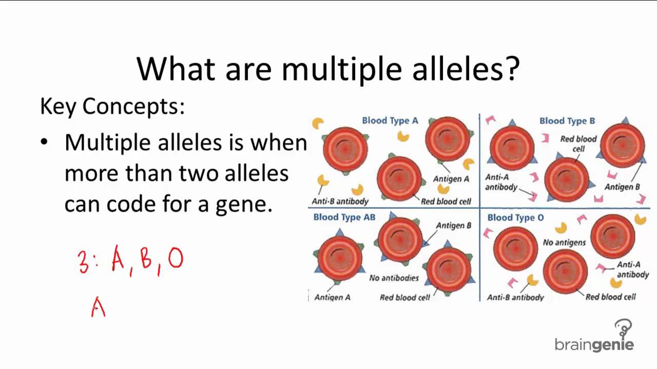
4.3.4 Describe ABO blood groups as an example of codominance and multiple alleles
When assigning alleles for codominance, the convention is to use a common letter to represent dominant and recessive and use superscripts to represent the different codominant alleles


4.3.5 Explain how the sex chromosomes control gender by referring to the inheritance of X and Y chromosomes in humans
Humans have 23 pairs of chromosomes for a total of 46 (excluding instances of aneuploidy)
The first 22 pairs are autosomes - each chromosome pair possesses the same genes and structural features
The 23rd pair of chromosomes are heterosomes (or sex chromosomes) and determine gender

4.3.6 State that some genes are present on the X chromosome and absent from the shorter Y chromosome in humans
The Y chromosome is much shorter than the X chromosome and contains only a few genes

4.3.7 Define sex linkage
Sex linkage refers to when a gene controlling a characteristic is found on a sex chromosome (and so we associate the trait with a predominant gender)

4.3.8 Describe the inheritance of colour blindness and hemophilia as examples of sex linkage.
Colour blindness and haemophilia are both examples of X-linked recessive conditions
The gene loci for these conditions are found on the non-homologous region of the X chromosome (they are not present of the Y chromosome)
As males only have one allele for this gene they cannot be a carrier for the condition
This means they have a higher frequency of being recessive and expressing the trait
Males will always inherit an X-linked recessive condition from their mother
Females will only inherit an X-linked recessive condition if they receive a recessive allele from both parents

4.3.9 State that a human female can be homozygous or heterozygous with respect to sex-linkage genes
As human females have two X chromosomes (and therefore two alleles for any given X-linked gene), they can be either homozygous or heterozygous
Males only have one X chromosome (and therefore only one allele) and are hemizygous
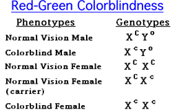
4.3.10 Explain that female carriers are heterozygous for X-linked recessive alleles.
An individual with a recessive allele for a disease condition that is masked by a normal dominant allele is said to be a carrier
Carrier are heterozygous and can potentially pass the trait on the next generation, but do not suffer from the defective condition themselves
Female can be carrier for X-linked recessive conditions because they have two X chromosomes - male (XY) cannot be carrier
Because a male only inherits an X chromosome from his mother, his chances of inheriting the disease condition from a carrier mother is greater

4.3.11 Predict the genotypic and phenotypic ratios of offspring of monohybrid crosses involving any of the above patterns of inheritance.
Monohybrid Cross:

Codominant Cross:

Sex Linkage Cross:

4.3.12 Deduce the genotypes and phenotypes of individuals in pedigree charts.
A pedigree is a chart of the genetic history of a family over several generations

Genotype: The allele combination of an organism
Phenotype: The characteristics of an organism (determined by a combination of genotype and environmental factors)
Dominant allele: An allele that has the same effect on the phenotype whether it is present in the homozygous or heterozygous state
Recessive allele: An allele that only has an effect on the phenotype when present in the homozygous state
Codominant allele: Pairs of alleles that both affect the phenotype when present in a heterozygous
Locus: The particular position on homologous chromosomes of a gene
Homozygous: Having two identical alleles of a gene
Heterozygous: Having two different alleles of a gene
Carrier: An individual that has one copy of a recessive allele that causes a genetic disease in individuals that are homozygous for this allele
Test cross: Testing a suspected heterozygote by crossing it with a known homozygous recessive
4.3.2 Determine the genotypes and phenotypes of the offspring of a monohybrid cross using a Punnett grid
A genetic cross is a means of determining the genetic characteristics of potenial offspring based on the genetic characteristics of the prospective parents
A monohybrid cross determines the allele combinations of offspring for one particular gene only
This is the general flow of how to show monohybrid crossing
4.3.3 State that some genes have more than two alleles (multiple alleles)
Some genes have more than two alleles for a given trait (e.g. the ABO blood group system)
- Share codominance (be expressed equally in the phenotype)
- Share incomplete dominance (neither is fully expressed in the phenotype, resulting in blending)
- Demonstrate a dominance order
4.3.4 Describe ABO blood groups as an example of codominance and multiple alleles
When assigning alleles for codominance, the convention is to use a common letter to represent dominant and recessive and use superscripts to represent the different codominant alleles
- I stands for immunoglobins (antigenic protein on blood cells)
- A and B stand for the codominant variants
4.3.5 Explain how the sex chromosomes control gender by referring to the inheritance of X and Y chromosomes in humans
Humans have 23 pairs of chromosomes for a total of 46 (excluding instances of aneuploidy)
The first 22 pairs are autosomes - each chromosome pair possesses the same genes and structural features
The 23rd pair of chromosomes are heterosomes (or sex chromosomes) and determine gender
- Females are XX - they possess two X chromosomes
- Males are XY - they possess one X chromosome and a much shorter Y chromosome
The Y chromosome contains the genes for developing male sex characteristic - hence the father is always responsible for determining gender
- If the male sperm contains the X chromosome the growing embryo will develop into a girl
- If the male sperm contains a Y chromosome the growing embryo will develop into a boy
- In all cases the female egg will contain an X chromosome (as the mother is XX)
Because the X and Y chromosome are of a different size, they cannot undergo crossing over / recombination during meiosis
This ensures that the gene responsible for gender always remains on the Y chromosome, meaning that there is always ~ 50% chance of a boy or girl
4.3.6 State that some genes are present on the X chromosome and absent from the shorter Y chromosome in humans
The Y chromosome is much shorter than the X chromosome and contains only a few genes
- Includes the SRY sex-determination gene and a few others (e.g. hairy ears gene)
The X chromosome is much longer and contains several genes not present on the Y chromosome
- Includes the genes for haemophilia and red-green colour blindness
In human females, only one of the X chromosomes remain active throughout life
- The other is packaged as heterochromatin to form a condensed Barr body
- This inactivation is random and individual to each cell, so heterozygous women will be a mosaic - expressing both alleles via different cells
4.3.7 Define sex linkage
Sex linkage refers to when a gene controlling a characteristic is found on a sex chromosome (and so we associate the trait with a predominant gender)
- Sex-linked conditions are usually X-linked, as very few genes exist on the shorter Y chromosomes
4.3.8 Describe the inheritance of colour blindness and hemophilia as examples of sex linkage.
Colour blindness and haemophilia are both examples of X-linked recessive conditions
The gene loci for these conditions are found on the non-homologous region of the X chromosome (they are not present of the Y chromosome)
As males only have one allele for this gene they cannot be a carrier for the condition
This means they have a higher frequency of being recessive and expressing the trait
Males will always inherit an X-linked recessive condition from their mother
Females will only inherit an X-linked recessive condition if they receive a recessive allele from both parents
4.3.9 State that a human female can be homozygous or heterozygous with respect to sex-linkage genes
As human females have two X chromosomes (and therefore two alleles for any given X-linked gene), they can be either homozygous or heterozygous
Males only have one X chromosome (and therefore only one allele) and are hemizygous
4.3.10 Explain that female carriers are heterozygous for X-linked recessive alleles.
An individual with a recessive allele for a disease condition that is masked by a normal dominant allele is said to be a carrier
Carrier are heterozygous and can potentially pass the trait on the next generation, but do not suffer from the defective condition themselves
Female can be carrier for X-linked recessive conditions because they have two X chromosomes - male (XY) cannot be carrier
Because a male only inherits an X chromosome from his mother, his chances of inheriting the disease condition from a carrier mother is greater
4.3.11 Predict the genotypic and phenotypic ratios of offspring of monohybrid crosses involving any of the above patterns of inheritance.
Monohybrid Cross:
Codominant Cross:
Sex Linkage Cross:
4.3.12 Deduce the genotypes and phenotypes of individuals in pedigree charts.
A pedigree is a chart of the genetic history of a family over several generations
- Males are represented as squares, while females are represented as circles
- Shaded symbols means an individual is affected by a condition, while an unshaded symbol means they are unaffected
- A horizontal line between a man and woman represent mating and resulting children are shown as offshoots to this line
Autosomal dominance
- All affected individuals must have at least one affected parent
- If two parents are unaffected, all offspring must be unaffected (homozygous recessive)
- If two parents are affected, they may have offspring who are unaffected (if parents are heterozygous)
Autosomal recessive
- If two parents shows a trait, all children must also the trait (homozygous recessive)
- An affected individual may have two normal parents (if parents are both heterozygous carriers)
X-linked recessive
- If a female shows the trait, so must all sons as well as her father
- The disorder is more common in males
訂閱:
文章 (Atom)