11.1.1 Describe the process of blood clotting
11.1.2 Outline the principle of challenge and response, clonal selection and memory cells as the basis of immunity
11.1.3 Define active and passive immunity
11.1.4 Explain antibody production
11.1.5 Describe the production of monoclonal antibodies and their use in diagnosis and in treatment
11.1.6 Explain the principle of vaccination
11.1.7 Discuss the benefits and dangers of vaccination
2014年6月9日 星期一

Topic 11: Human health and physiology
Topic 11 of the IB HL Biology syllabus is the Genetics. IBO recommends to spend 17 hours on this topic.
This topic has 4 sub-chapters: "Defense against infectious disease", "Muscles and movement", "The kidney" and "Reproduction". Each are separated with numerical values in order of mentioned.
These are all HL syllabus statements, it is recommended to bring a Casio Graphical Calculator instead of Texas.
Topic 6.6: Reproduction
6.6.1 Draw and label diagrams of the adult male and female reproductive systems
6.6.2 Outline the role of hormones in the menstrual cycle, including FSH (follicle stimulating hormones), LH (luteinizing hormone), estrogen and progesterone
6.6.3 Annotate a graph showing hormone levels in the menstrual cycle, illustrating the relationship between changes in hormone level and ovulation, menstruation and thickening of the endometrium
6.6.4 List three roles of testosterone in males
6.6.5 Outline the process of in vitro fertilization (IVF)
6.6.6 Discuss the ethical issues associated with IVF
Topic 6.5: Nerves, hormones and homeostasis
6.5.1 State that the nervous system consists of the central nervous system (CNS) and peripheral nerves, and is composed of cells called neurons that can carry rapid electrical impulses
6.5.2 Draw and label a diagram of the structure of a motor neuron
6.5.3 State that nerve impulses are conducted from recpetors to the CNS by sensory neurons, within the CNS by relay neurons, and from the CNS to effectors by motor neurons
6.5.4 Define resting potential and action potential (depolarization and repolarization)
6.5.5 Explain how a nerve impulse passes along a non-myelinated neuron
6.5.6 Explain the principles of synaptic transmission
6.5.7 State that the endocrine system consists of glands that release hormones that are transported in the blood
6.5.8 State that homeostasis involves maintaining the internal environment between limits, including blood pH, carbon dioxide concentration, blood glucose concentration, body temperature and water balance
6.5.9 Explain that homeostasis involves monitoring levels of variables and correcting changes in levels by negative feedback mechanism
6.5.10 Explain the control of body temperature, including the transfer of heat in blood, and the roles of the hypothalamus, sweat glands, skin arterioles and shivering
6.5.11 Explain the control of blood glucose concentration, including the roles of glucagon, insulin and a and b cells in the pancreatic islets
6.5.12 Distinguish between type I and type II diabetes
Topic 6.4: Gas exchange
6.4.1 Distinguish between ventilation, gas exchange and cell respiration
6.4.2 Explain the need for a ventilation system
6.4.3 Describe the features of alveoli that adapt them to gas exchange
6.4.4 Draw and label a diagram of the ventilation system, including trachea, lungs, bronchi, bronchioles and alveoli
6.4.5 Explain the mechanism of ventilation of the lungs in terms of volume and pressure changes caused by the internal and external intercostal muscles, the diaphragm and abdominal muscles
Topic 6.3: Defense against infectious disease
6.3.1 Define pathogen
A pathogen is an organism or virus that causes a disease. Most, but not all, are microorganisms.

6.3.2 Explain why antibiotics are effective against bacteria but not against viruses
Antibiotics are naturally occuring substances that slow down or kill microorganisms. They are obtained mainly from fungi or bacteria, and are substances which such organisms manufacture in their natural habitats.
Most antibiotics are so extremely effective at disrupting bacterial metabolism that whole populations are quickly suppressed. A key process here is the formation and laying down of the new dividing wall. Bacterial cells have a rigid wall containing giant, complex, molecules of amino-sugars and peptide units (polymers) rather than the polysaccharides of plant cell walls.

Virus on the other hand, are not living cells and have no metabolism of their own to be interfered with. Viruses reproduce using metabolic pathways in their host cell that are not affected by antibiotics. Antibiotics cannot be used to prevent viral diseases.

6.3.3 Outline the role of skin and mucous membranes in defense against pathogens
6.3.4 Outline how phagocytes leucocytes ingest pathogens in the blood and in blood tissues
6.3.5 Distinguish between antigens and antibodies
6.3.6 Explain antibody production
6.3.7 Outline the effects of HIV on the immune system
6.3.8 Discuss the cause, transmission and social implications of AIDs
Topic 6.2: The transport system
6.2.1 Draw and label a diagram of the heart showing the four chambers, associated blood vessels, valves and the route of blood through the heart

6.2.2 State that the coronary arteries supply heart muscle with oxygen and nutrients
The heart is a muscle itself, so note that the walls of the heart are supplied with oxygenated blood via coronary arteries. These arteries and the capillaries they serve, deliver to the muscle fibres of the heart the oxygen and the nutrients essential for the pumping action.

6.2.3 Explain the action of the heart in terms of collecting blood, pumping blood, and opening and closing of valves
The cavity of the heart is divided into four chambers, with those on the right side of the heart completely separate from those on the left. The two upper chambers are called the artia and these receive blood into the heart.
The lower chambers are thick-walled ventricles, with the muscular wall of the left ventricle much thicker than that of the right ventricle. However, the volumes of the right and left sides (the quantities of blood they contain) are identical. The ventricles pump blood out of the heart.

The valves of the heart prevent backflow of the blood, thereby maintaining the direction of flow through the heart. The artio-ventricular valves are large valves, positioned to prevent backflow from ventricles to artia. The edges of these valves are supported by tendons anchored to the muscle walls of the ventricles below. A different type of valve separates the ventricles from the pulmonary artery and the aorta. These are called the semi-lunar valves.

6.2.4 Outline the control of the heartbeat in terms of myogenic muscle contraction, the role of the pacemaker, nerves, the medulla of the brain and epinephrine (adrenaline)
The heart normally beats about 75 times per minute - approximately 0.8 seconds per beat. In each beat, the heart muscle contracts strongly, followed by a period of relaxation.
The atrium contacts pushing blood past the bicuspid valve into the ventricle, Atrium muscle relax. Ventricle muscles contract, causing blood pressure to close the bicuspid valve and open the semilunar valves, forcing blood into the aorta. Ventricle and atrium muscle relax, while the pressure of blood in the aorta causes the semilunar valves to shut. Blood flows into the atrium and opens the bicuspid valve as it starts to flow into the ventricle.
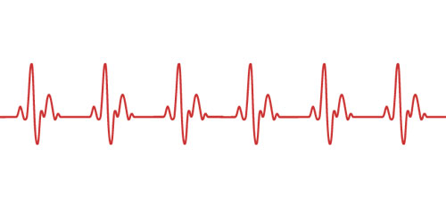 The heart beats rhythmically throughout life, without rest, apart from the momentary relaxation between beats. Even more remarkably, the origin of each beat is within the heart itself - we say the heart beat is myogenic in origin.
Beats originate in a strucuture in the muscle of the wall of the right atrium, called the pacemaker. Special muscle fibre radiate out from the pacemaker, conducting the impulse to the muscles of both artia, triggering contraction. Then a second excitation is passed onto the ventricles causing a ventricular contraction.

The heart receives impulses from a control centre in the hindbrain (medulla), via two nerves. One nerve, when stimulated, triggers speeding up of the heart rate, and the other nerve triggers a slowing down of the heart. Since these nerves have opposite effect, they are antagonistic.

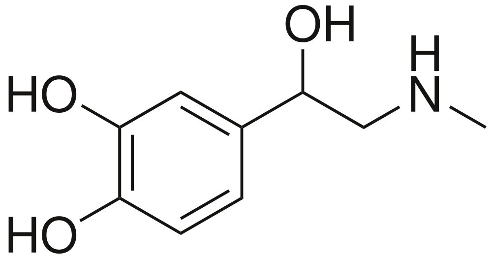
The hormone adrenaline is secreted by the adrenal glands and carried in the blood, causes the pacemaker to increase the heart rate.

6.2.5 Explain the relationship between the structure and function of arteries, capillaries and veins
There are three types of vessels in the circulation system.
Arteries
- Carry blood away from the heart
- Carry blood at high pressures
- Have a narrow lumen
- Have a thick middle layer to help the pulse flow
- Have a outer layer to prevent rupture
Capillaries
- Are a fine networks of tiny tubes linking arteries and veins.
- Involved with material and gas exchange with the surrounding body tissues
- Blood pressure is relatively low
- Small diametre
- Single cell to allow easier diffusion
- Contains pores to aid the transport of materials
Veins
- Carry blood to the heart
- Carry blood at low pressure
- Very wide lumen
- Have valves to prevent blooding pooling
- Thin surrounding tissue as blood do not go in rhythmic pulse.

6.2.6 State that blood is composed of plasma, erythrocytes, leucocytes (phagocytes and lymphocytes) and platelets
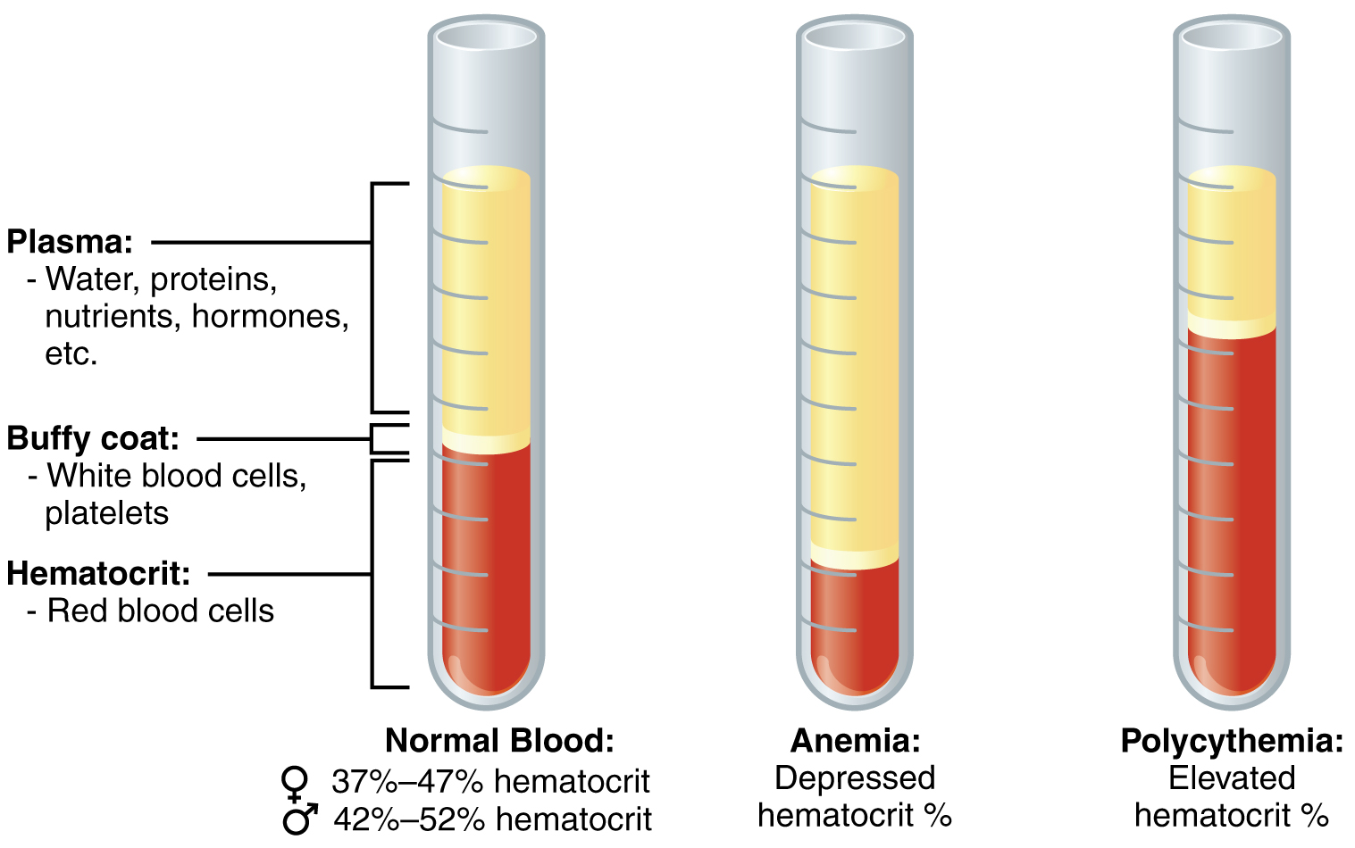
Blood is a special tissue consisting of a liquid medium called plasma in which are suspended red cells or erythrocytes, white cells or luecocytes, and platelets. The plasma is the medium for exchange of substances between cells and tissues, the erythrocytes are involved in transport of respiratory gases.

6.2.7 State that the following are transported by the blood: nutrients, oxygen, carbon dioxide, hormones, antibodies, urea and heat
The blood circulation has roles in the body's defense against diseases as well as being the all-important transport system of the body. Nutrients from digestion, oxygen and carbon dioxide, urea, hormones and antibodies are all transported. In the tissues of the body, exchange between the blood and cells of the tissues occurs from the capillaries, the walls of which are permeable and highly "leaky".
Tissue respiration - gas exchange
Hydration - water to all the tissues
Nutrition - Nutrients (sugars, amino acids, lipids, vitamins) and inorganic ions to all cells.
Excretion - Waste product urea to kidneys
Development and co-ordination - Hormones from endocrine glands to target organs
Temperature regulation - distribution of heat
Defense against disease - Antibodies are circulated in the blood system


6.2.2 State that the coronary arteries supply heart muscle with oxygen and nutrients
The heart is a muscle itself, so note that the walls of the heart are supplied with oxygenated blood via coronary arteries. These arteries and the capillaries they serve, deliver to the muscle fibres of the heart the oxygen and the nutrients essential for the pumping action.
6.2.3 Explain the action of the heart in terms of collecting blood, pumping blood, and opening and closing of valves
The cavity of the heart is divided into four chambers, with those on the right side of the heart completely separate from those on the left. The two upper chambers are called the artia and these receive blood into the heart.
The lower chambers are thick-walled ventricles, with the muscular wall of the left ventricle much thicker than that of the right ventricle. However, the volumes of the right and left sides (the quantities of blood they contain) are identical. The ventricles pump blood out of the heart.
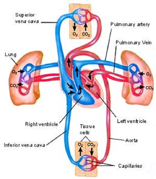
The valves of the heart prevent backflow of the blood, thereby maintaining the direction of flow through the heart. The artio-ventricular valves are large valves, positioned to prevent backflow from ventricles to artia. The edges of these valves are supported by tendons anchored to the muscle walls of the ventricles below. A different type of valve separates the ventricles from the pulmonary artery and the aorta. These are called the semi-lunar valves.

6.2.4 Outline the control of the heartbeat in terms of myogenic muscle contraction, the role of the pacemaker, nerves, the medulla of the brain and epinephrine (adrenaline)
The heart normally beats about 75 times per minute - approximately 0.8 seconds per beat. In each beat, the heart muscle contracts strongly, followed by a period of relaxation.
The atrium contacts pushing blood past the bicuspid valve into the ventricle, Atrium muscle relax. Ventricle muscles contract, causing blood pressure to close the bicuspid valve and open the semilunar valves, forcing blood into the aorta. Ventricle and atrium muscle relax, while the pressure of blood in the aorta causes the semilunar valves to shut. Blood flows into the atrium and opens the bicuspid valve as it starts to flow into the ventricle.
Beats originate in a strucuture in the muscle of the wall of the right atrium, called the pacemaker. Special muscle fibre radiate out from the pacemaker, conducting the impulse to the muscles of both artia, triggering contraction. Then a second excitation is passed onto the ventricles causing a ventricular contraction.

The heart receives impulses from a control centre in the hindbrain (medulla), via two nerves. One nerve, when stimulated, triggers speeding up of the heart rate, and the other nerve triggers a slowing down of the heart. Since these nerves have opposite effect, they are antagonistic.

The hormone adrenaline is secreted by the adrenal glands and carried in the blood, causes the pacemaker to increase the heart rate.
6.2.5 Explain the relationship between the structure and function of arteries, capillaries and veins
There are three types of vessels in the circulation system.
Arteries
- Carry blood away from the heart
- Carry blood at high pressures
- Have a narrow lumen
- Have a thick middle layer to help the pulse flow
- Have a outer layer to prevent rupture
Capillaries
- Are a fine networks of tiny tubes linking arteries and veins.
- Involved with material and gas exchange with the surrounding body tissues
- Blood pressure is relatively low
- Small diametre
- Single cell to allow easier diffusion
- Contains pores to aid the transport of materials
Veins
- Carry blood to the heart
- Carry blood at low pressure
- Very wide lumen
- Have valves to prevent blooding pooling
- Thin surrounding tissue as blood do not go in rhythmic pulse.

6.2.6 State that blood is composed of plasma, erythrocytes, leucocytes (phagocytes and lymphocytes) and platelets
Blood is a special tissue consisting of a liquid medium called plasma in which are suspended red cells or erythrocytes, white cells or luecocytes, and platelets. The plasma is the medium for exchange of substances between cells and tissues, the erythrocytes are involved in transport of respiratory gases.
6.2.7 State that the following are transported by the blood: nutrients, oxygen, carbon dioxide, hormones, antibodies, urea and heat
The blood circulation has roles in the body's defense against diseases as well as being the all-important transport system of the body. Nutrients from digestion, oxygen and carbon dioxide, urea, hormones and antibodies are all transported. In the tissues of the body, exchange between the blood and cells of the tissues occurs from the capillaries, the walls of which are permeable and highly "leaky".
Tissue respiration - gas exchange
Hydration - water to all the tissues
Nutrition - Nutrients (sugars, amino acids, lipids, vitamins) and inorganic ions to all cells.
Excretion - Waste product urea to kidneys
Development and co-ordination - Hormones from endocrine glands to target organs
Temperature regulation - distribution of heat
Defense against disease - Antibodies are circulated in the blood system

訂閱:
文章 (Atom)