
6.2.2 State that the coronary arteries supply heart muscle with oxygen and nutrients
The heart is a muscle itself, so note that the walls of the heart are supplied with oxygenated blood via coronary arteries. These arteries and the capillaries they serve, deliver to the muscle fibres of the heart the oxygen and the nutrients essential for the pumping action.

6.2.3 Explain the action of the heart in terms of collecting blood, pumping blood, and opening and closing of valves
The cavity of the heart is divided into four chambers, with those on the right side of the heart completely separate from those on the left. The two upper chambers are called the artia and these receive blood into the heart.
The lower chambers are thick-walled ventricles, with the muscular wall of the left ventricle much thicker than that of the right ventricle. However, the volumes of the right and left sides (the quantities of blood they contain) are identical. The ventricles pump blood out of the heart.
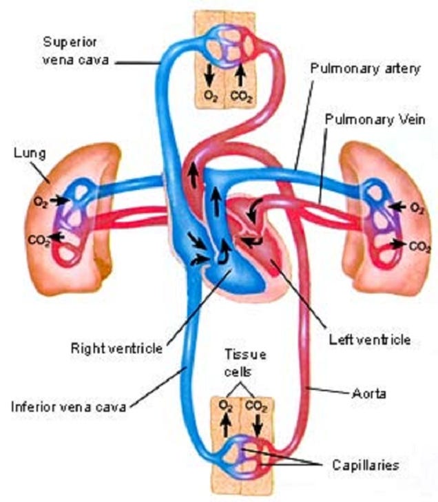
The valves of the heart prevent backflow of the blood, thereby maintaining the direction of flow through the heart. The artio-ventricular valves are large valves, positioned to prevent backflow from ventricles to artia. The edges of these valves are supported by tendons anchored to the muscle walls of the ventricles below. A different type of valve separates the ventricles from the pulmonary artery and the aorta. These are called the semi-lunar valves.

6.2.4 Outline the control of the heartbeat in terms of myogenic muscle contraction, the role of the pacemaker, nerves, the medulla of the brain and epinephrine (adrenaline)
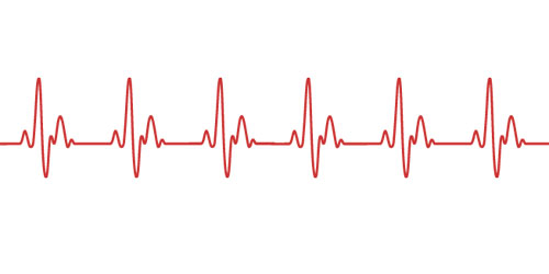
The heart normally beats about 75 times per minute - approximately 0.8 seconds per beat. In each beat, the heart muscle contracts strongly, followed by a period of relaxation.
The atrium contacts pushing blood past the bicuspid valve into the ventricle, Atrium muscle relax. Ventricle muscles contract, causing blood pressure to close the bicuspid valve and open the semilunar valves, forcing blood into the aorta. Ventricle and atrium muscle relax, while the pressure of blood in the aorta causes the semilunar valves to shut. Blood flows into the atrium and opens the bicuspid valve as it starts to flow into the ventricle.
Beats originate in a strucuture in the muscle of the wall of the right atrium, called the pacemaker. Special muscle fibre radiate out from the pacemaker, conducting the impulse to the muscles of both artia, triggering contraction. Then a second excitation is passed onto the ventricles causing a ventricular contraction.

The heart receives impulses from a control centre in the hindbrain (medulla), via two nerves. One nerve, when stimulated, triggers speeding up of the heart rate, and the other nerve triggers a slowing down of the heart. Since these nerves have opposite effect, they are antagonistic.

The hormone adrenaline is secreted by the adrenal glands and carried in the blood, causes the pacemaker to increase the heart rate.
6.2.5 Explain the relationship between the structure and function of arteries, capillaries and veins
There are three types of vessels in the circulation system.
Arteries
- Carry blood away from the heart
- Carry blood at high pressures
- Have a narrow lumen
- Have a thick middle layer to help the pulse flow
- Have a outer layer to prevent rupture
Capillaries
- Are a fine networks of tiny tubes linking arteries and veins.
- Involved with material and gas exchange with the surrounding body tissues
- Blood pressure is relatively low
- Small diametre
- Single cell to allow easier diffusion
- Contains pores to aid the transport of materials
Veins
- Carry blood to the heart
- Carry blood at low pressure
- Very wide lumen
- Have valves to prevent blooding pooling
- Thin surrounding tissue as blood do not go in rhythmic pulse.

6.2.6 State that blood is composed of plasma, erythrocytes, leucocytes (phagocytes and lymphocytes) and platelets
Blood is a special tissue consisting of a liquid medium called plasma in which are suspended red cells or erythrocytes, white cells or luecocytes, and platelets. The plasma is the medium for exchange of substances between cells and tissues, the erythrocytes are involved in transport of respiratory gases.

6.2.7 State that the following are transported by the blood: nutrients, oxygen, carbon dioxide, hormones, antibodies, urea and heat
The blood circulation has roles in the body's defense against diseases as well as being the all-important transport system of the body. Nutrients from digestion, oxygen and carbon dioxide, urea, hormones and antibodies are all transported. In the tissues of the body, exchange between the blood and cells of the tissues occurs from the capillaries, the walls of which are permeable and highly "leaky".
Tissue respiration - gas exchange
Hydration - water to all the tissues
Nutrition - Nutrients (sugars, amino acids, lipids, vitamins) and inorganic ions to all cells.
Excretion - Waste product urea to kidneys
Development and co-ordination - Hormones from endocrine glands to target organs
Temperature regulation - distribution of heat
Defense against disease - Antibodies are circulated in the blood system

沒有留言:
張貼留言