2014年3月28日 星期五

Topic 4.3: Theoretical genetics

4.3.1 Define genotype, phenotype, dominant allele, recessive allele, codominant alleles, locus, homozygous, heterozygous, carrier and test cross

Genotype: The allele combination of an organism

Phenotype: The characteristics of an organism (determined by a combination of genotype and environmental factors)

Dominant allele: An allele that has the same effect on the phenotype whether it is present in the homozygous or heterozygous state

Recessive allele: An allele that only has an effect on the phenotype when present in the homozygous state

Codominant allele: Pairs of alleles that both affect the phenotype when present in a heterozygous

Locus: The particular position on homologous chromosomes of a gene

Homozygous: Having two identical alleles of a gene

Heterozygous: Having two different alleles of a gene

Carrier: An individual that has one copy of a recessive allele that causes a genetic disease in individuals that are homozygous for this allele

Test cross: Testing a suspected heterozygote by crossing it with a known homozygous recessive


4.3.2 Determine the genotypes and phenotypes of the offspring of a monohybrid cross using a Punnett grid

A genetic cross is a means of determining the genetic characteristics of potenial offspring based on the genetic characteristics of the prospective parents

A monohybrid cross determines the allele combinations of offspring for one particular gene only


This is the general flow of how to show monohybrid crossing


4.3.3 State that some genes have more than two alleles (multiple alleles)

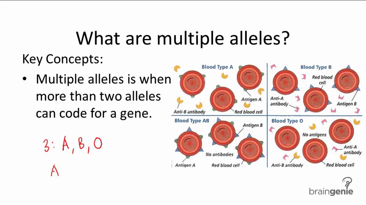
Some genes have more than two alleles for a given trait (e.g. the ABO blood group system)

  • Share codominance (be expressed equally in the phenotype)
  • Share incomplete dominance (neither is fully expressed in the phenotype, resulting in blending)
  • Demonstrate a dominance order


4.3.4 Describe ABO blood groups as an example of codominance and multiple alleles

When assigning alleles for codominance, the convention is to use a common letter to represent dominant and recessive and use superscripts to represent the different codominant alleles

  • I stands for immunoglobins (antigenic protein on blood cells)
  • A and B stand for the codominant variants


4.3.5 Explain how the sex chromosomes control gender by referring to the inheritance of X and Y chromosomes in humans

Humans have 23 pairs of chromosomes for a total of 46 (excluding instances of aneuploidy)

The first 22 pairs are autosomes - each chromosome pair possesses the same genes and structural features

The 23rd pair of chromosomes are heterosomes (or sex chromosomes) and determine gender

  • Females are XX - they possess two X chromosomes
  • Males are XY - they possess one X chromosome and a much shorter Y chromosome
The Y chromosome contains the genes for developing male sex characteristic - hence the father is always responsible for determining gender
  • If the male sperm contains the X chromosome the growing embryo will develop into a girl
  • If the male sperm contains a Y chromosome the growing embryo will develop into a boy
  • In all cases the female egg will contain an X chromosome (as the mother is XX)
Because the X and Y chromosome are of a different size, they cannot undergo crossing over / recombination during meiosis

This ensures that the gene responsible for gender always remains on the Y chromosome, meaning that there is always ~ 50% chance of a boy or girl


4.3.6 State that some genes are present on the X chromosome and absent from the shorter Y chromosome in humans

The Y chromosome is much shorter than the X chromosome and contains only a few genes

  • Includes the SRY sex-determination gene and a few others (e.g. hairy ears gene)
The X chromosome is much longer and contains several genes not present on the Y chromosome
  • Includes the genes for haemophilia and red-green colour blindness

In human females, only one of the X chromosomes remain active throughout life
  • The other is packaged as heterochromatin to form a condensed Barr body
  • This inactivation is random and individual to each cell, so heterozygous women will be a mosaic - expressing both alleles via different cells 


4.3.7 Define sex linkage

Sex linkage refers to when a gene controlling a characteristic is found on a sex chromosome (and so we associate the trait with a predominant gender)

  • Sex-linked conditions are usually X-linked, as very few genes exist on the shorter Y chromosomes


4.3.8 Describe the inheritance of colour blindness and hemophilia as examples of sex linkage.

Colour blindness and haemophilia are both examples of X-linked recessive conditions

The gene loci for these conditions are found on the non-homologous region of the X chromosome (they are not present of the Y chromosome)

As males only have one allele for this gene they cannot be a carrier for the condition

This means they have a higher frequency of being recessive and expressing the trait

Males will always inherit an X-linked recessive condition from their mother

Females will only inherit an X-linked recessive condition if they receive a recessive allele from both parents


4.3.9 State that a human female can be homozygous or heterozygous with respect to sex-linkage genes

As human females have two X chromosomes (and therefore two alleles for any given X-linked gene), they can be either homozygous or heterozygous

Males only have one X chromosome (and therefore only one allele) and are hemizygous
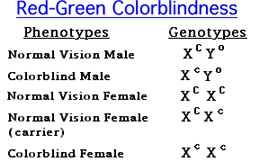


4.3.10 Explain that female carriers are heterozygous for X-linked recessive alleles.

An individual with a recessive allele for a disease condition that is masked by a normal dominant allele is said to be a carrier

Carrier are heterozygous and can potentially pass the trait on the next generation, but do not suffer from the defective condition themselves

Female can be carrier for X-linked recessive conditions because they have two X chromosomes - male (XY) cannot be carrier

Because a male only inherits an X chromosome from his mother, his chances of inheriting the disease condition from a carrier mother is greater


4.3.11 Predict the genotypic and phenotypic ratios of offspring of monohybrid crosses involving any of the above patterns of inheritance.

Monohybrid Cross:


Codominant Cross:

Sex Linkage Cross:


4.3.12 Deduce the genotypes and phenotypes of individuals in pedigree charts.

A pedigree is a chart of the genetic history of a family over several generations

  • Males are represented as squares, while females are represented as circles
  • Shaded symbols means an individual is affected by a condition, while an unshaded symbol means they are unaffected
  • A horizontal line between a man and woman represent mating and resulting children are shown as offshoots to this line
Autosomal dominance
  • All affected individuals must have at least one affected parent
  • If two parents are unaffected, all offspring must be unaffected (homozygous recessive)
  • If two parents are affected, they may have offspring who are unaffected (if parents are heterozygous)
Autosomal recessive
  • If two parents shows a trait, all children must also the trait (homozygous recessive)
  • An affected individual may have two normal parents (if parents are both heterozygous carriers)
X-linked recessive
  • If a female shows the trait, so must all sons as well as her father
  • The disorder is more common in males


Topic 4.2: Meiosis

4.2.1 State that meiosis is a reduction division of a diploid nucleus to form haploid nuclei

Meiosis is a reduction division of a diploid nucleus to form haploid nuclei

Meiosis makes gametes (sex cells), which are produced in the reproductive organs (gonads).


4.2.2 Define homologous chromosomes

Homologous chromosomes resemble each other in structure. They occur in diploid cell, contain the same sequence of genes, but have come from different parents. They have the same genes at the same loci positions


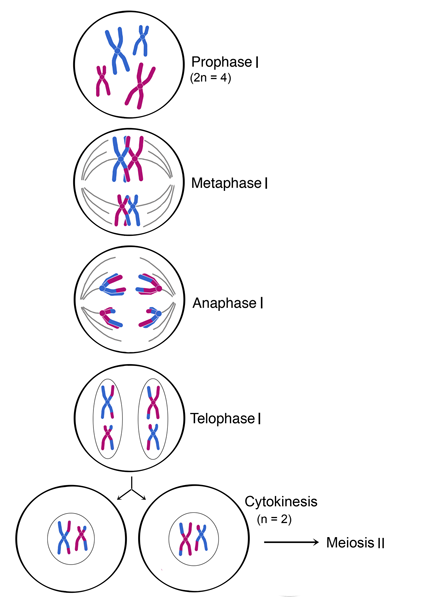
4.2.3 Outline the process of meiosis, including pairing of homologous chromosomes and crossing over, followed by two divisions, which results in four haploid cells

The process of meiosis involves two divisions, both of which follow the same basic stages as mitosis (prophase, metaphase, anaphase and telophase)

Meiosis is preceded by interphase, which includes the replication of DNA (S phase) to create chromosomes with genetically identical sister chromatids


Meiosis I

Homologous chromosomes must first pair up in order to be sorted into separate haploid daughter cells

In prophase I, homologous chromosomes undergo a process called synapsis, whereby homologous chromosomes pair up to forma  bivalent (or tetrad)

  • The homologous chromosomes are held together at points called chiasma (singular: chiasmata)
  • Crossing over of genetic material between non-sister chromatids can occur at these points, resulting in new gene combination (recombination)
  • Crossing over and independent assortment are the process which provides the genetic variation

The remainder of meiosis I involves separating the homologous chromosomes into separate daughter cells

  • In meetaphase I, the homologous chromosomes pairs line up along the equator of the cell
  • In anaphase I, the homologous chromosomes split apart and move to opposite poles
  • In telophase I, the cell splits into two haploid daughter cells as cytokinesis happens concurrently


Meiosis II

In meiosis II, the sister chromatids are divided into separate cells

  • In prophase II, spindle fibre reform and reconnect to the chromosomes
  • In metaphase II, the chromosomes line up along the equator of the cell
  • In anaphase II, the sister chromatids split apart and move to opposite poles
  • In telophase II, the cell splits in two as cytokinesis happens concurrently.


Because sister chromatids may no longer be genetically identical as a result of potenial recombination, the process of meiosis results in the formation of four generally distinct haploid daughter cells


4.2.4 Explain that non-disjunction can lead to changes in chromosome number, illustrated by reference to Down syndrome (trisomy 21)

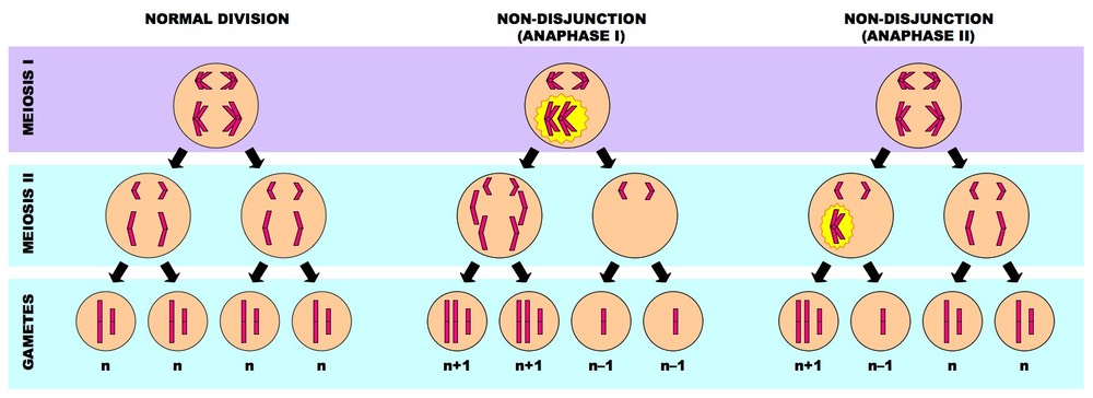
Non-disjunction refers to the chromosomes failing to separate correctly, resulting in gametes with one extra, or one missing chromosome (aneuploidy)

The failure of the chromosomes to separate may either occur via:

  • Failure of homologous to separate during Anaphase I (resulting in four affected daughter cells)
  • Failure of sister chromatids to separate during Anaphase II (resulting in two affected daughter cells)


Individuals with down syndrome has a trisomy 21 (three chromosomes 21)


4.2.5 State that, in karyotyping, chromosomes are arranged in pairs according to their size and structure

Karyotyping is a visual profile of all the chromosomes in a cell

The chromosomes are arranged into homologous pairs and displayed according to their structural characteristics


4.2.6 State that karyotyping is performed using cells collected by chorionic villus sampling or amniocentesis, for pre-natal diagnosis of chromosome abnormalities

Pre-natal karyotyping is often used to:

  • Determine the gender of an unborn child (via identification of sex chromosomes)
  • Test for chromosomal abnormalities (e.g. aneuploidies resulting from non-disjunction)
Amniocentesis 
  • A needle is inserted through the abdominal wall, into the amniotic cavity in the uterus, and a sample of amniotic fluid containing foetal cells is taken
  • It can be done at ~ 16th week of pregnancy, with a slight chance of miscarriage (~0.5%)
Chorionic Villus Sampling
  • A tube is inserted through the cervix and a tiny sample of the chronic villi (contains foetal cells) from the placenta is taken
  • It can be done at ~ 11th week of pregnancy, with a slight risk of inducing miscarriage (~ 1%)


4.2.7 Analyse a human karyotype to determine gender and whether non-disjunction has occured.

Inside every cell in the human body, there are 46 chromosomes (except for red blood cells and haploid gametes)

Males have a X,Y chromosome while the female has X,X

Non-disjunction during gamete formation can lead to individuals with an abnormal number of chromosomes


Topic 4.1: Chromosomes, genes, alleles and mutations

4.1.1 State that eukaryote chromosomes are made of DNA and proteins

An eukaryotic cell's chromosomes are made up of DNA and proteins. They are structured around 8 histone balls to form a nucleosome. This is shown in the image below.


A collection of nucleosomes are called a chromatin. It is important to not that, DNA is a nucleic acid. It is different from protein even if part of the DNA is made up of nitrogenous bases. Hence it is incorrect to state that it is wrong to write that histones are made up of protein.


4.1.2 Define gene, allele and genome

Gene: A heritable factor that controls a specific characteristic, consisting of a length of DNA occupying a particular position on a chromosome (loci)

Allele: One specific form of a gene, differing from other alleles by one or a few bases only and occupying the same locus as other alleles of the gene.

Genome: The whole of the genetic information of an organism


4.1.3 Define gene mutation

Gene Mutation: A change in the nucleotide sequence of a section of DNA coding for a particular feature.


4.1.4 Explain the consequence of a base substitution mutation in relation to the processes of transcription and translation, using the example of sickle-cell anemia.

A base substitution mutation is the change of a single base in a sequence of DNA

This resulting in a change to a single mRNA codon during transcription

In sickle cell anemia, the DNA sequence for the beta chain of haemoglobin is changed from GAG to GTG

This changes the mRNA codon (GAG to GUG), resulting in a single amino acid change (Glu to Val)

The amino acid change alters the structure of the haemoglobin causing it to form insoluble strands

This causes the red blood cells to adopt a sickle shape


The insoluble haemoglobin can't effectively carry oxygen, causing individuals to feel constantly tired

The sickle cells may accumulate in the capillaries and form clots, blocking blood supply to organs

Also causes anemia (low RBC count), as sickle cells are destroyed more rapidly than normal cells

Sickle cell anemia occurs in individuals who have two copies of the codominant "sickle cell" allele

Heterozygous individuals have increased resistance to malaria


Topic 4: Genetics

Topic 4 of the IB HL Biology syllabus is the Genetics. IBO recommends to spend 15 hours on this topic.

This topic has 4 sub-chapters: "Chromosomes, genes, alleles and mutations", "Meiosis", "Theoretical genetic" and "Genetic engineering and biotechnology". Each are separated with numerical values in order of mentioned.

These are all SL syllabus statements, it is recommended to bring a Casio Graphical Calculator instead of Texas.

2014年3月25日 星期二

Topic 8.2: Photosynthesis

8.2.1 Draw and label a diagram showing the structure of a chloroplast as seen in electron micrograph


8.2.2 State that photosynthesis consists of light-dependent and light-independent reactions

Photosynthesis is a two step process which consists of light dependent (converts light energy into chemical energy) and light independent variable (uses chemical energy to make organic molecules)


8.2.3 Explain the light-dependent reactions

The light dependent reaction occurs on the thylakoid membrane

Chlorophyll in both photosystem I and II absorbs light, which triggers the release of high energy electrons (photoactivation)

  • The electrons from photosystem II pass along a series of carriers
  • The electrons lost from photosystem II are replaced by electrons generated by the photolysis of water with oxygen as by-product
  • Hydrogen pump carriers use the excited electrons to pump hydrogen ions over into the thylakoid compartment, the electrons is then passed on to photosystem I
  • Photosystem I re-excites the electrons and then passed on to NADP+ reductase
  • The electrons then binds with the NADPH + H+ and is transported somewhere else.
  • ATP synthase uses the concentration gradient to produce ATP from ADP and Pi


8.2.4 Explain photophosphorylation in terms of chemiosmosis

Photophosphorylation is simply phosphorylation but required light energy.

As the electrons cycle through the electron transport chain located on the thylakoid membrane, they lose energy. The energy is used to pump H+ ions into the thylakoid compartment to create a concentration gradient. The H+ ions return via ATP synthase.

This process is called chemiomosis


8.2.5 Explain the light-independent reactions

Calvin cycle occurs in the stroma and uses ATP and NADPH + H+ produced by the light dependent reaction. There are three main steps which include carbon fixation, reduction and regeneration of RuBP.

Carbon fixation:

  • Enzyme RuBisCo catalyses the attachment of carbon dioxide to the 5 carbon compound ribulose bisphosphate (RuBP)
  • The unstable 6 carbon compound that is formed immediately breaks down into two 3 carbon molecules called glycerate-3-phosphate (GP)

Reduction:
  • ATP is used to put phosphate onto the GP
  • NADPH + H+ reduces the compound by giving a hydrogen.
  • This forms G3P

Regeneration
  • For every six molecules of G3P produced, only one could be used to form half a sugar
  • Th remaining G3P is used to restock RuBP in a reaction that require ATP
  • With RuBP regenerated, the plant will use the cycle multiple times and construct long chains of sugars.


8.2.6 Explain the relationship between the structure of the chloroplast and its function


8.2.7 Explain the relationship between the action spectrum and the absorption spectrum of photosynthetic pigments in green plants.

Pigments require light as a source of energy.

The absorption spectrum indicates the wavelength of light absorbed by each pigment. The action spectrum indicates the rate of photosynthesis for each wavelength.

There are strong relationships between the two as it shows both the peak and the valleys in the graph.


8.2.8 Explain the concept of limiting factors in photosynthesis, with reference to light intensity, temperature and concentration of carbon dioxide

The law of limiting factor states that when a chemical process depends on more than one essential condition to become favorable, its rate will be limited by the factors that is nearest its minimum value.

Light intensity

  • Light is required for the light dependent reactions (photoactivation of chlorophyll and photolysis of water molecules)
  • Low light intensities results in insufficient production of ATP and NADPH + H+
Temperature
  • Primarily affect light-independent reaction as it requires more collisions and enzymes
  • High temperatures could damage the enzymes and cause the prohibition of enzymes from occuring
Concentration of Carbon Dioxide
  • Carbon dioxide is required for the light independent reaction to occur (carbon fixation of RuBP by RuBisCo)
  • At low levels, oxygen will over take the enzyme RuBisCo creating toxic gas instead.


Topic 8.1: Cell Respiration

8.1.1 State that oxidation involves the loss of electrons from an element, whereas reduction involves a gain of electrons; and that oxidation frequently involves gaining oxygen or losing hydrogen, whereas reduction frequently involves losing oxygen or gaining hydrogen.


OILRIG - Oxidation Is Loss, Reduction Is Gain
ELMO - Electron Loss Means Oxidation


8.1.2 Outline the process of glycolysis, including phosphorylation, lysis, oxidation and ATP formation

Glycolysis takes place in the cytoplasm.


The process phosphorylation simple oxidizes the glucose present with phosphate groups. It attaches itself to both ends of the glucose for it to become Hexose Biphosphate. This process requires ATP for the phosphate group to attach itself.


Lysis is the stage when the hexose biphosphate molecule becomes too unstable and breaks down into two triose phosphate.


This is the oxidation/ATP formation step. Each of the triose phosphate is oxidized to become a pyruvate molecule. With the addition of one hydrogen, it is passed on and reduce one NAD+ to NADH. Each Triose phosphate adds a phosphate group to the ADP reducing it to ATP. Note: Each Triose phosphate releases enough energy to form two ADP to ATP.


This is the full sequence of glycolysis in short.


8.1.3 Draw and label a diagram showing the structure of a mitochondrion as seen in electron micrographs

This is the structure of a mitochondrion.


It is important to remember that Mitochondrion has a double membrane thus the existence of Cristae is to increase inner membrane surface area. Important for electron transport chain.


8.1.4 Explain aerobic respiration, including the link reaction, the Krebs cycle, the role of NADH + H+, the electron transport chain and the role of oxygen

There are 3 more stages in the aerobic respiration. This includes Link reaction, the Krebs cycle and the electron transport chain.

Link Reaction: Pyruvate is decarboxylated to become Acetyl CoA


The link reaction allows for pyruvate and Co-enzyme A to join together to form a complex. The carbon is decarboxylated (removal of carbon) as carbon dioxide. The remains forms Acetyl CoA. The NAD+ is reduced to NADH + H+.


Kreb's cycle: Oxidative decarboxylation of the acetyl group.


Acetyl CoA joins with a 4 carbon group to form citrate. CoA is then released to transport more pyruvate molecules. The C6 compound formed is called citric acid.


Citric acid is oxidatively decarbonxylated. A C5 group is now formed. The carbon is released as carbon dioxide. NAD+ is reduced to NADH + H+


The C5 grouped is also oxidatively decarbonxylated, forming a C4 group. The carbon is released as carbon dioxide. NAD+ is reduced to NADH + H+.


The final stage in the cycle requires the C4 group to regenerate back to its original form and accept acetyl CoA. This reduces NAD to NADH + H+ and FAD to FADH + H+. ADP also uses this energy to bind with a phosphate group to make ATP.

This is the whole process and shows how it is linked to the electron transport chain.


The hydrogen carriers (NADH + H+ and FADH + H+) provide electrons to the electron transport chain on the inner mitochondrial membrane. As the electrons cycle through the chain, they lose energy to translocate the hydrogen ions to the intermembrane space (creating a gradient). The hydrogen ions return to the matrix through ATP synthase mass producing ATP. Oxygen acts as the final electron acceptors for the electron transport chain to allow new electrons enter the chain. Oxygen combines with the hydrogen ions in the matrix to form water molecules.


8.1.5 Explain oxidative phosphorylation in terms of chemiosmosis

This is the electron transport chain.


  • Oxidative phosphorylation describes the production of ATP from oxidised hydrogen carriers
  • When electrons are donated to the electron transport chain, they lose energy as they are passed between submissive carrier molecules.
  • The energy is used to translocate H+ ions from the matrix to the intermembrane space against the concentration gradient.
  • The build up of H+ ions creates an electrochemical gradient, or proton motive force (PMF)
  • The protons return to the matrix via a transmembrane exnzyme called ATP synthase.
  • As the hydrogen ions return, they release energy which is used to produce ATP (from ADP + Pi)
  • This process is called chemiomosis and occurs in the cristae
  • The hydrogen ions and electrons bind with oxygen to form water molecules. 


8.1.6 Explain the relationship between the structure of the mitochondrian and its function

There are four structures in the mitochondrion which function to improve the reactions happening. Which include: Inner membrane, external double membrane, matrix and inter-membrane space

Inner membrane: The double folded inner membrane space forms cristae, this allows an increase of ATP synthesis thus more ATP produce
External double membrane: Contains appropriate proteins to allow the transport of molecules
Intermembrane space: Small space to increase the gradient difference.
Matrix: The right pH levels for the reaction and enzymes to work at optimal rate.


Topic 8: Cellular respiration and photosynthesis

Topic 8 of the IB HL Biology syllabus is the Cellular respiration and photosynthesis. IBO recommends to spend 10 hours on this topic.

This topic has 2 sub-chapters: "Cell respiration" and "Photosynthesis". Each are separated with numerical values in order of mentioned.

These are all HL syllabus statements, it is recommended to bring a Casio Graphical Calculator instead of Texas.